Matlab Generated Endothelial Cell Alignment Distribution
Top and Bottom Gel
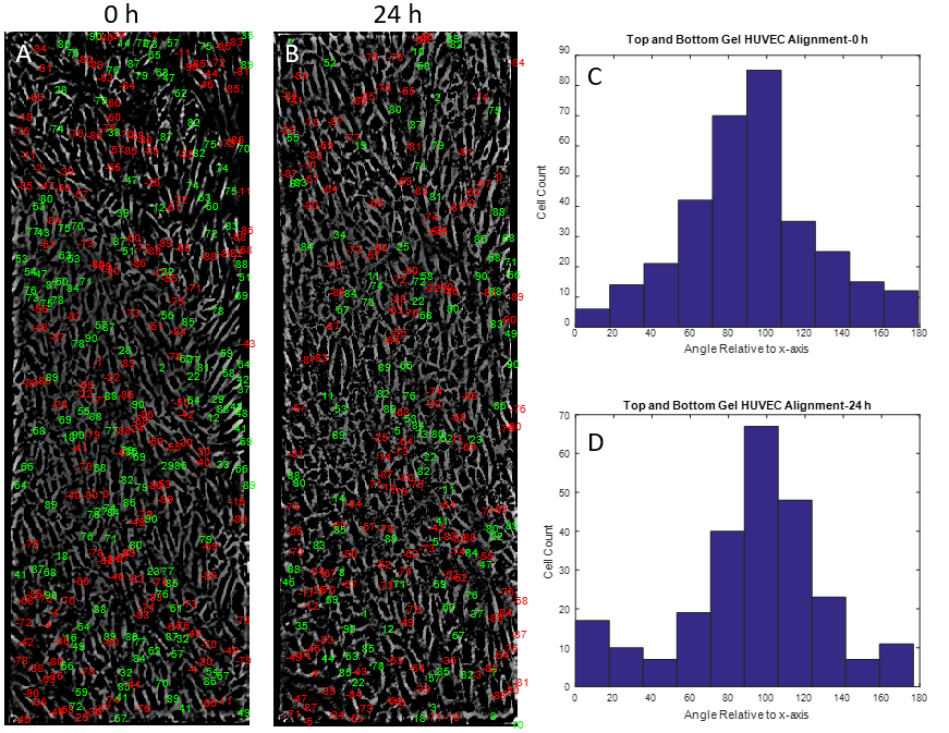
Figure 1. Matlab based alignment semi-quantification of HUVECs seeded on top of a collagen I gel. (A) Matlab output of major axis angle (relative to the x-axis) of HUVECs before flow set on top of cell image. (B) HUVEC angle of alignment relative to x-axis after 24 h flow. (C) Histogram plot of A, angles changed to reflect y-axis alignment. (D) Histogram plot of B.
No Gel
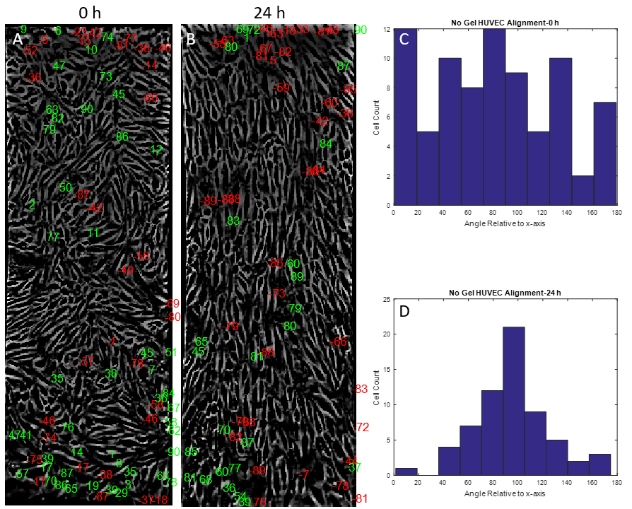
Figure 2. Matlab based alignment semi-quantification of HUVECs seeded directly on the NPN chip. (A) Matlab output of major axis angle (relative to the x-axis) of HUVECs before flow set on top of cell image. (B) HUVEC angle of alignment relative to x-axis after 24 h flow. (C) Histogram plot of A, angles changed to reflect y-axis alignment. (D) Histogram plot of B.
Bottom Gel
Run 1
Figure 3. Matlab based alignment semi-quantification of HUVECs seeded directly on the NPN chip with collagen I gel on the reverse side. (A) Matlab output of major axis angle (relative to the x-axis) of HUVECs before flow set on top of cell image. (B) HUVEC angle of alignment relative to x-axis after 24 h flow. (C) Histogram plot of A, angles changed to reflect y-axis alignment. (D) Histogram plot of B.
Run 2
Figure 4. Matlab based alignment semi-quantification of HUVECs seeded directly on the NPN chip with collagen I gel on the reverse side (RUN 2). (A) Matlab output of major axis angle (relative to the x-axis) of HUVECs before flow set on top of cell image. (B) HUVEC angle of alignment relative to x-axis after 24 h flow. (C) Histogram plot of A, angles changed to reflect y-axis alignment. (D) Histogram plot of B.
Statistical Analysis
The next step in this data analysis is to perform some form of statistical analysis. Comparing real alignment data to a set of randomly generated alignments may be a good place to start. Also, comparing the frequency distributions of the before flow and after flow alignment histograms may provide some level of significance.
Example Shear Calculations with 1um Polystyrene Beads
Matlab Video Processing
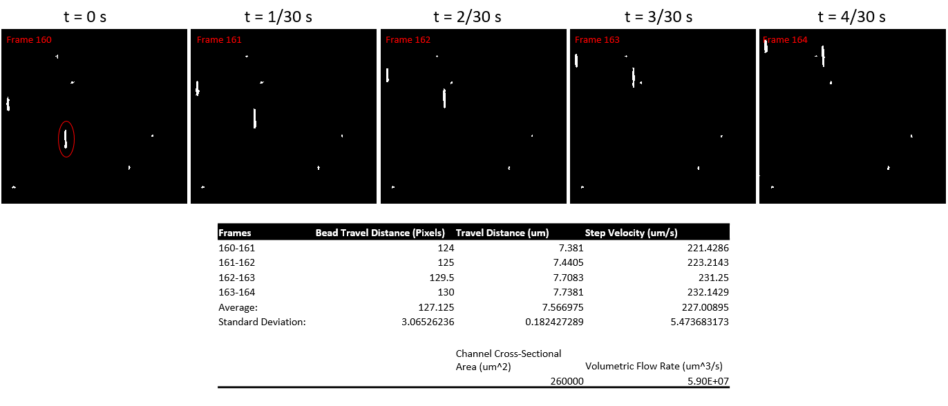
Using matlab, the video seen above was converted to gray scale frames, which were then converted to binary images (Figure 5). Matlab then tracks the center of the particle over 5 frames and calculates a distance traveled in pixels, distance traveled in um, and velocity in um/s.
Figure 5. (Top) Images showing particle trajectory over 4/30 s. Each picture represents a frame of a video shot at 30 fps. The particle circled in red was used to obtain velocity. (Bottom) Data from matlab analysis on particle velocity.
Using the formula for shear flow through a slit:
τ=6Qμ/(wh^2 )
I calculated a shear at the surface of the gel of about 0.093 dyn/cm^2. The expected shear at this level is around 6.5 dyn/cm^2.
Barrett had discovered that growing endothelial cells on pnc-si or tranwells causes vacuoles to form. The vacuoles on pnc-si formed only on the free standing areas of the membrane where there are open pores. The background on pnc-si chips showed negligible vacuole formation. The density of vacuoles on pnc-si was almost two fold higher than…
The x-values are based on TEM images of the last populations of gold nanoparticles (I think. I can’t remember immediately when the fridge died on us). It’s probably worth re-doing that with the new gold vials. There’s still some work to be done. In the 50 nm NPN, 5 nm gold was only done once,…
The physical properties of the extracellular matrix such as stiffness, viscoelasticity, fiber diameter, and chemical composition are well known as important modulators of cell behavior in vivo. Therefore, over the past decade, there has been a major emphasis on characterizing the properties of the in vivo ECM and also engineered matrices in vitro. In this context, properties such…
I wanted to attempt pressurized and diffusion separations of brain extract using the new movable wafers. I tried pressure cell separations of 1:20 BBEC (clarified brain extract) at 2psi for 40 min. The flow rates were better than the standards I was previously using – this could be due to the little bit of glycerol…
The main difficulty with creating a simulation of the single channel dialysis chip is that the membrane is so very thin (20 nm). This is especially true when compared to its length of 10 mm but also when compared to the channel depth and permeate well (which in our case is actually a beaker of…