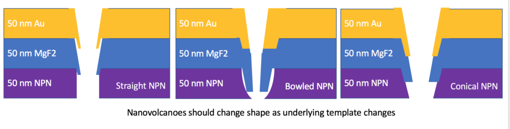
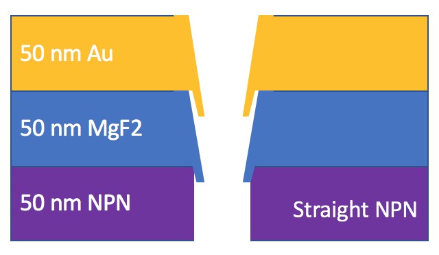
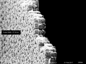
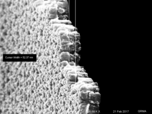
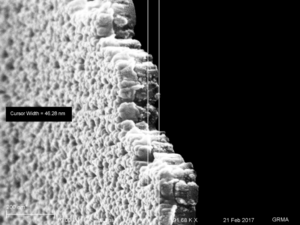
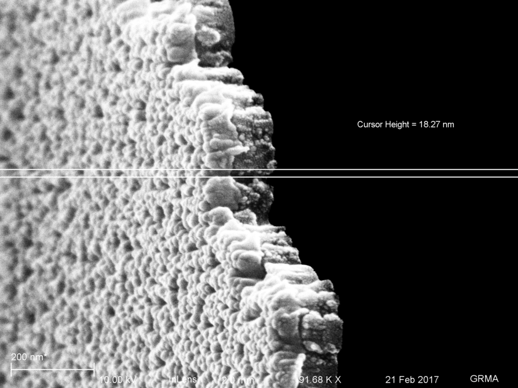
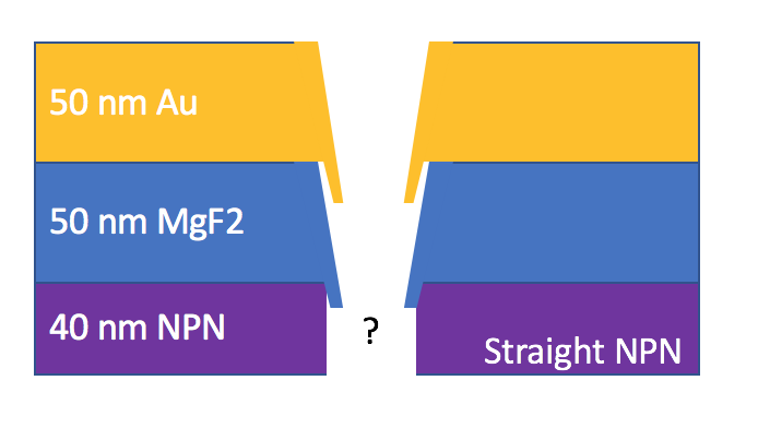
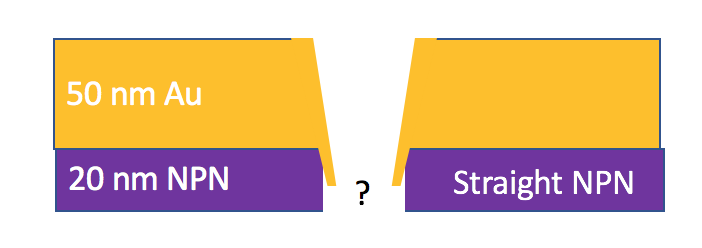
This post is a continuation of the work in Partial Etch RIE Blows out NPN Pores and MgF2 Nanomembrane Fabrication on partially etched NPN substrates. We believe the underlying NPN template influences the shape of our SERS active nanostructures and therefore the SERS properties. In this post, we’ll take a look at some of the structures resulting from partially etching the NPN template, thinning and broadening the template, which will change the shape of the structures that are fabricated upon them.
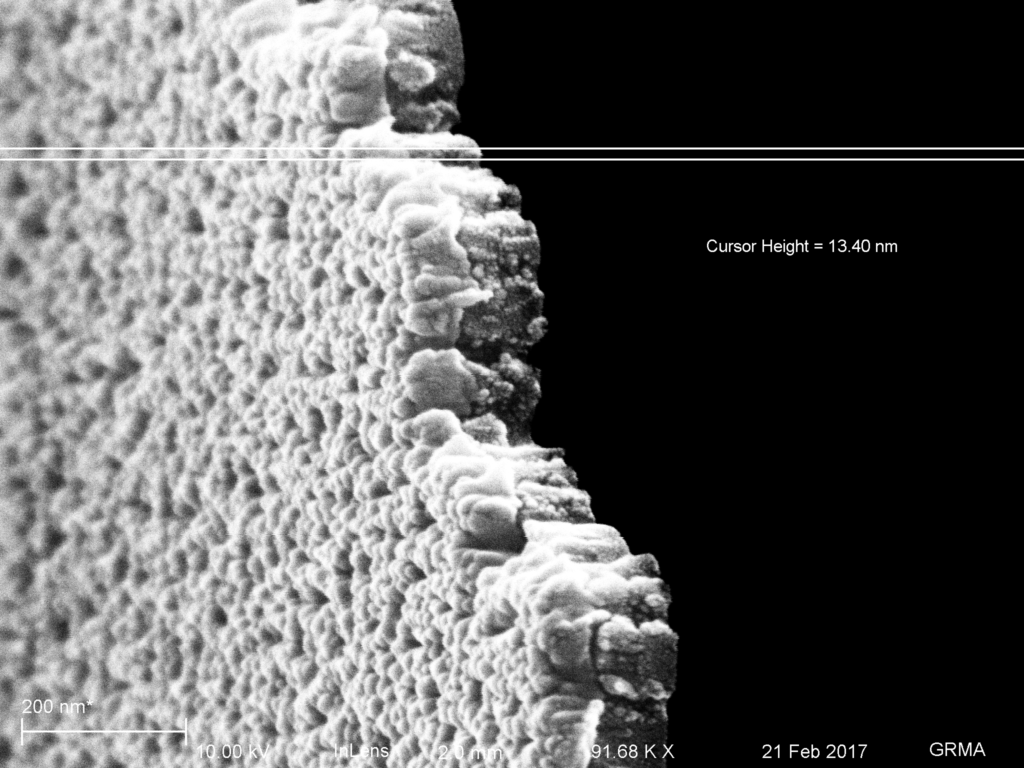
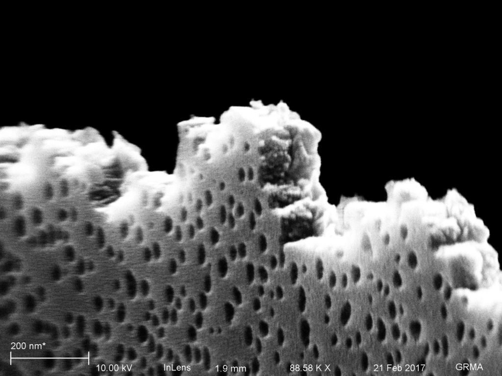
After the fabrication, each of these membranes were fractured and then coated with 8-9 nm of platinum. I will probably use less next time, as it was occluding my crossections.
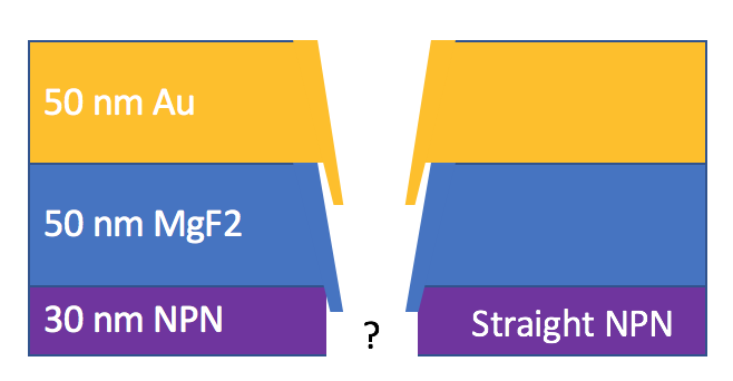
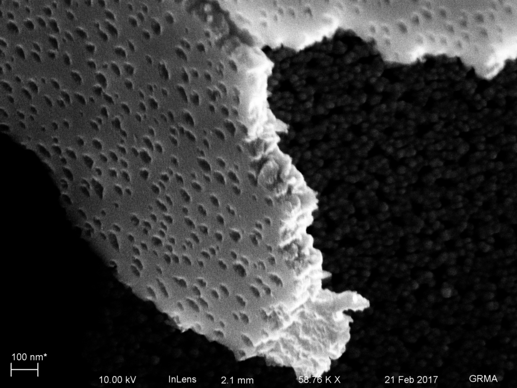
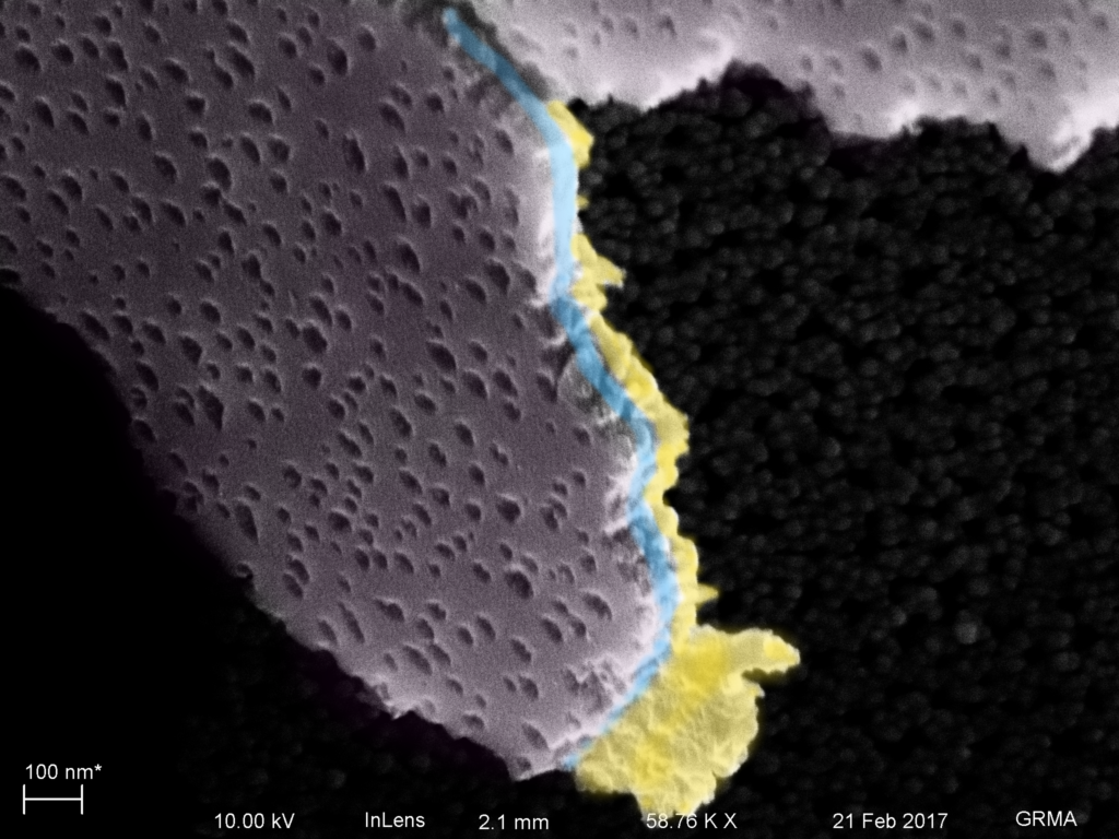
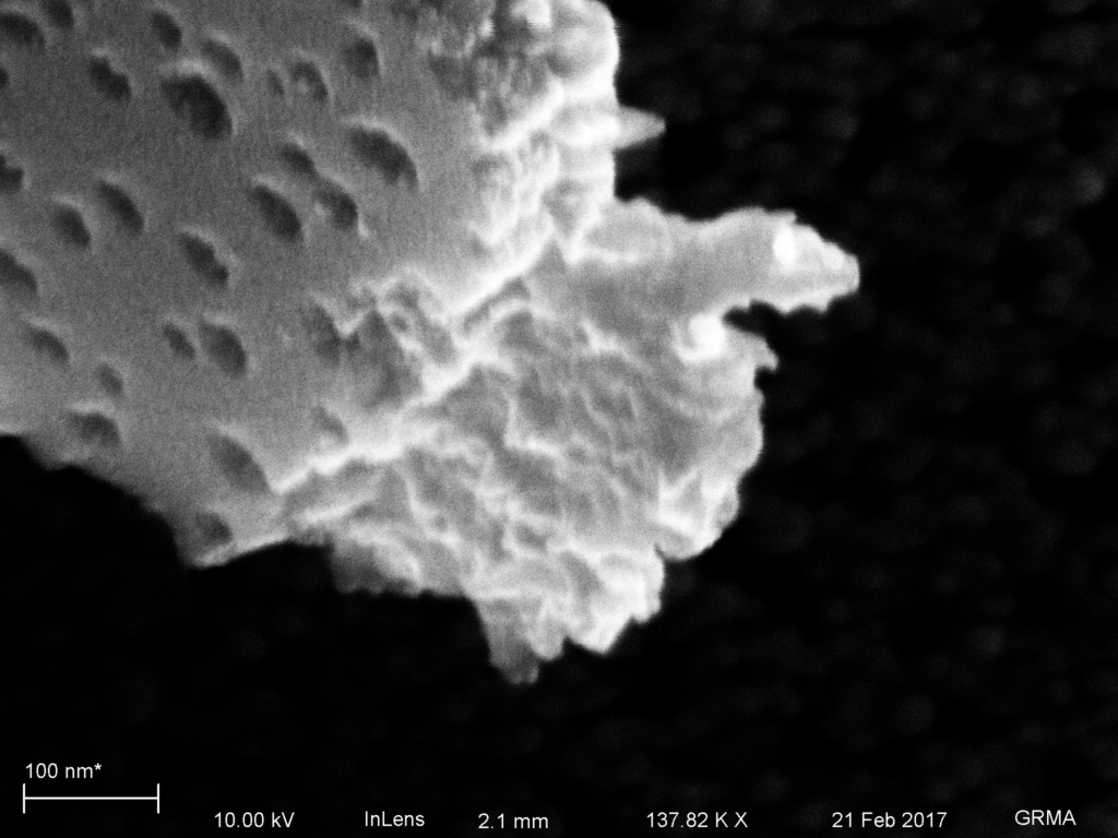
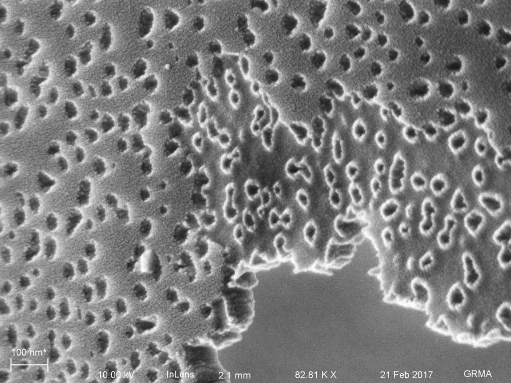
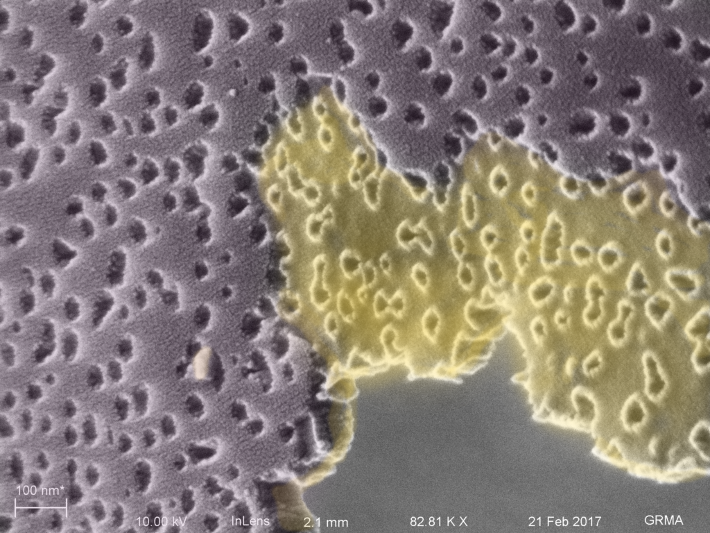
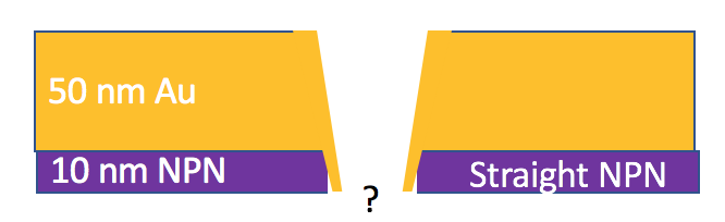
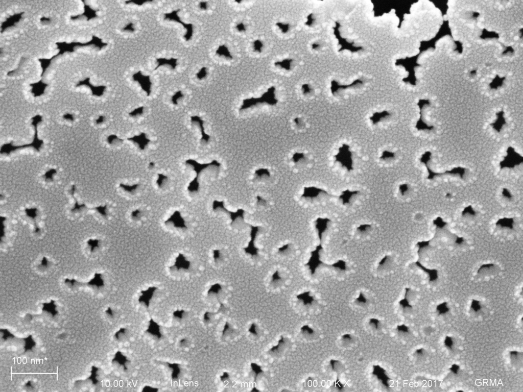
10 s etch viewed from the back. Fractures through the NPN are pretty straight, not conical.
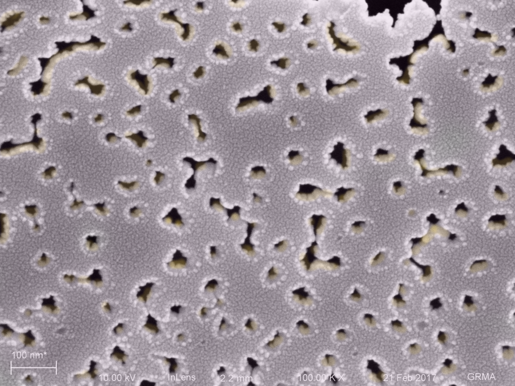
Colorized 10 s etch
20s etch 1236 NPN + MgF2 + Au
We’re not certain to what degree does the partial etched membrane have conical features.
20s etch. The NPN template is thinning. and we can see that the gold has some interesting pointed shapes when peeled away.20 s colorized
30s etch 1236 NPN + Au
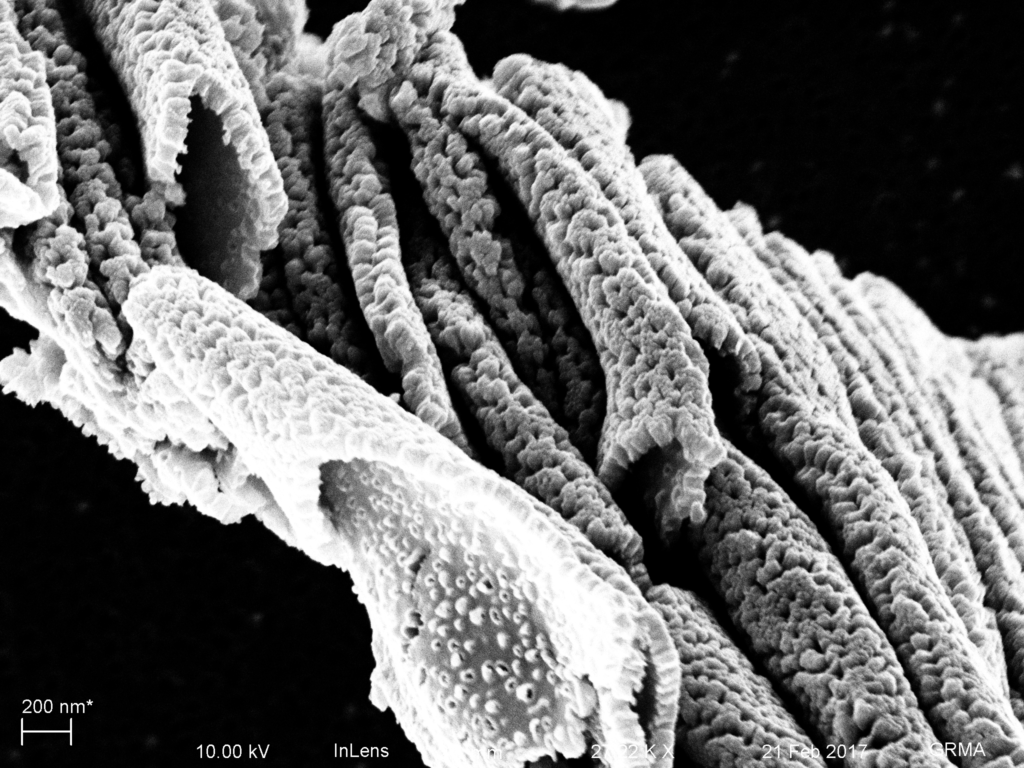
Since the much thinned NPN template cannot survive the MgF2 evaporation stress, we coat gold on the thinned NPN directly.30 s etch. Thinned NPN reveals gold volcanoes.30 s etch colorized.Scraping up the gold in the bulk region shows that volcanoes are formed with some porosity. Internally, they may be more pinched than the shape of the volcano indicates.
40 s etch 1236 NPN + Au
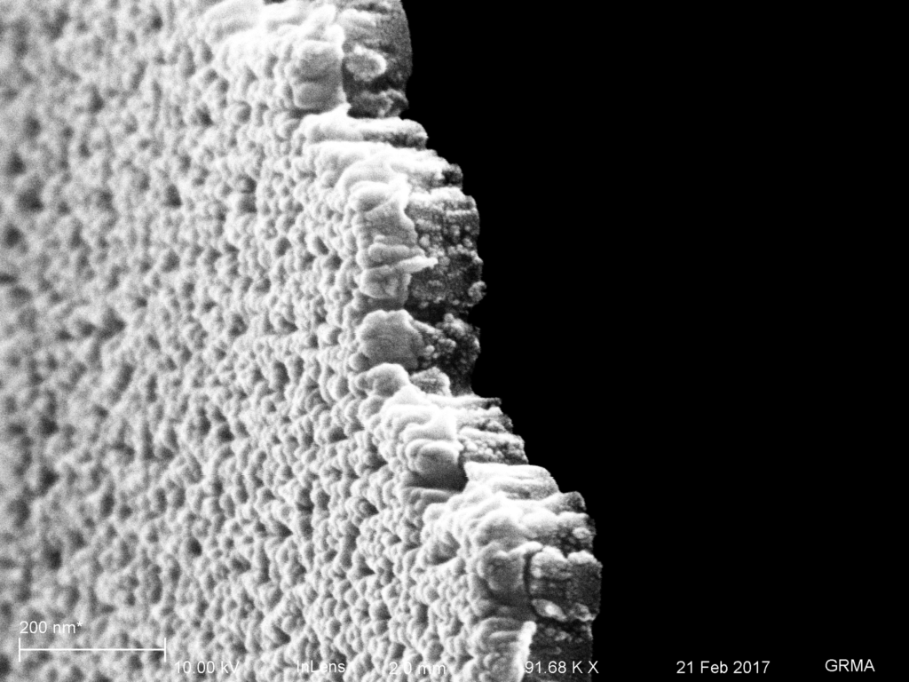
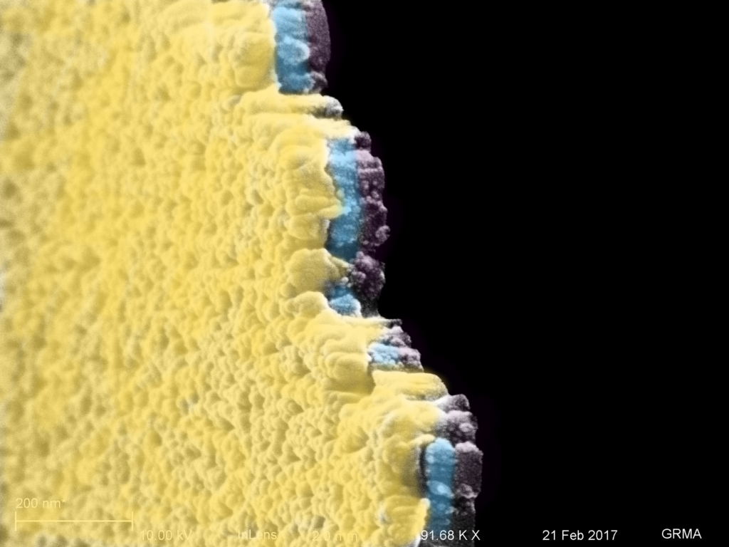
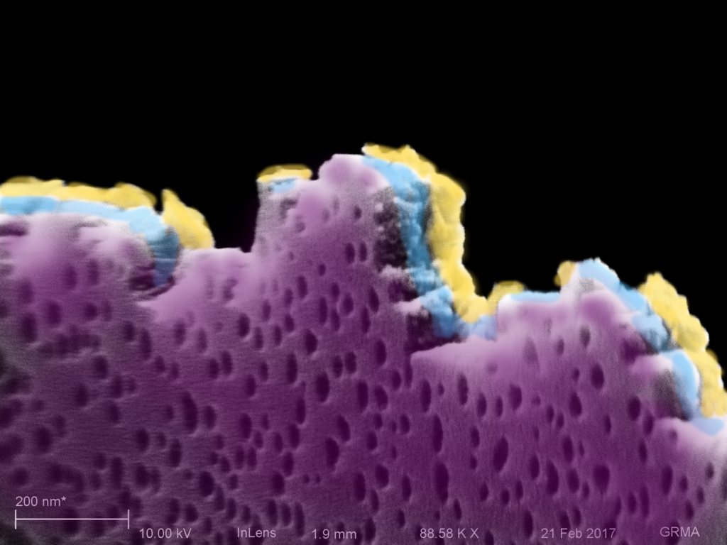
40 s etch. Viewed from the backside, it is clear that the features of the NPN have changed, as more pores have merged together. Thin bits of gold are viewable through the pores, as it comprises most of the film thickness.
40 s colorized
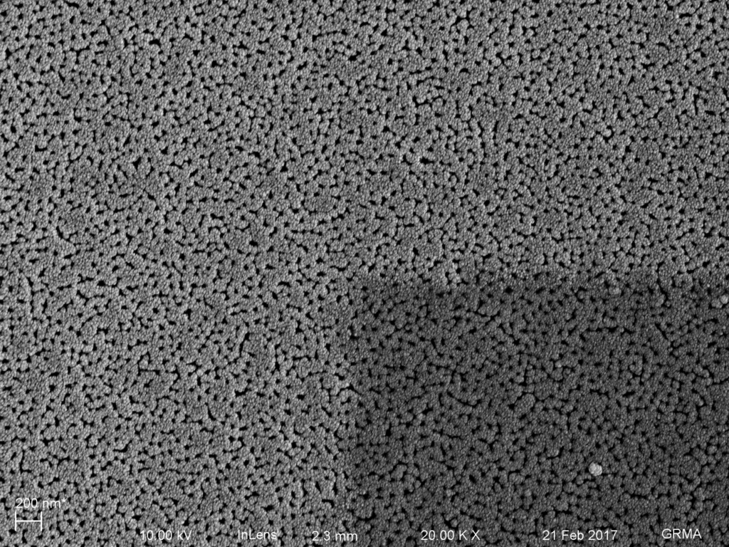
Aerial Image shows a contiguous membrane into the freestanding region (darker).
As Aim 1 of my thesis involves using theoretical models to be able to predict particle capture, it is important for me to fully understand how each model system behaves when compared to experimental results. One of the most important of these systems is the pressure drop across the membrane in our devices. I have…
My previous post outlined the results of a EQ format separations using membranes from wafer 319. I have followed up that experiment using the same membranes, but in a diffusion based separation. I separated 2uL of 9.3 mg/mL clarified brain extract using a reservoir of 60uL. I allowed the diffusion process to occur overnight, and…
I reran the 100K Microcon membranes using bovine albumin. This time I ran a set of four fresh membranes with DI water for a preliminary trial. Once complete I used two of the same four membranes with a 1mg/mL solution of bovine albumin in PBS to test the effects of protein; the other two membranes…
Just in case you missed this one: www.nature.com.ezp.lib.rochester.edu/nnano/journal/v2/n11/full/nnano.2007.354.html This article makes the the previous papers seem mundane. It has inspired me though – maybe we can create a computer simulation that will demonstrate that pnc-Si membranes will dance and sing songs under the correct conditions. I will leave the choice of genre to the programmer…
To follow up on my recent posts about HUVEC YAP localization after 24 and 72 hours (https://trace-bmps.org/blog/data/2016/10/19/yaptaz-passage-one-staining and https://trace-bmps.org/blog/data/2016/10/11/yaptaz-staining-on-membranes-and-nonporous-sio2) , we conducted the same study using Adipose Derived Stem Cells. Due to the fact that these are mesenchymal stem cells, we thought that their expression of YAP might be a bit different due to the…
I’ve been taking lots of zetapotential measurements with my current setup. Serendipitously, it appears that the more I use the electrodes, the more stable my zetapotential measurements become. Because the system seems well behaved now, I decided to move on to a more difficult problem. Sarah and I made some nanofluidic transistors a few weeks back,…