Kidney Week 2017
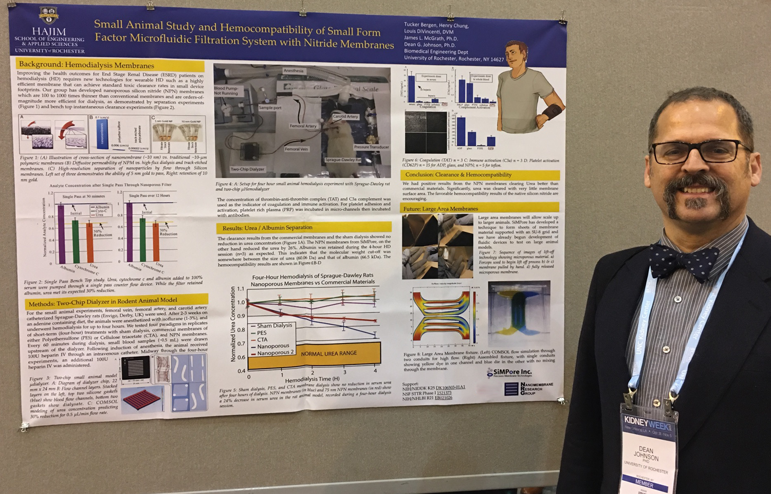
I had Five objectives for my trip to this conference.
1. Meet with my K25 Program Director, Tracy Rankin
Its is always a good idea to meet face-to-face when you can. Program directors have a lot of grants in their portfolios and it is good to make contact and introduce yourself so when you need something from them, they can associate the request with a person. Tracy remembered that my first annual report was well written and covered all the tasks I was supposed to have accomplished, whether I accomplished them or not. I also met two other Program Directors from NIH NIDDK, Deborah Hoshizaki (Kidney, Urologic, and Hematologic Diseases) and someone from the Intramural Administrative Management Branch. (I have his name on a piece of paper somewhere). Deborah recommended finding two or three people on the RBK consortium (see “A new Kidney to Call My Own” below) and invite them to UR for a seminar where they would give 30 min talks, this is to expand the potential scope of my research with an R01 in mind.
2. Meet with the Pittsburgh member of my advisory panel, Dr. John Kellum.
I updated Dr. Kellum on my K25 progress and laid out plans to finish the in vitro bench work and the final, I hope, round of rat studies with PEGylated chips. He is well satisfied with my progress and plans. We also discussed sheep vs pig for large animal studies. The main reason people use pigs if because the immune system more closely models that of the human immune system. We’ll have to decide if we’re going to investigate this in our four hour studies. He did want to make sure we’re going to put our results in a format that nephrologists will recognize, URR, kT/V etc. and that we measure things like thrombin count, complement activation in the animal.
3. Meet with members of my research community. This turned our to be Shuvo Roy, Bill Fissell, Victor Gura, who are all working on implantable or wearable RRT devices; Frank Hurst and Iwen Wu from the FDA. I’ve been running into the same group of people at KHI, ASAIO, and Kidney Week over the past two years. Frank, who works with the KHI wants to add me to the membrane advisory panel, once they form a membrane advisory panel.
4. Present my Poster, which has some of the data we’ll be including in the paper we’re working on. This includes the small animal model and the previous in situ data. I did have a lot of interest, and spent the better part of two hours (1.5 probably) talking non-stop to explain our membranes and what they can do.
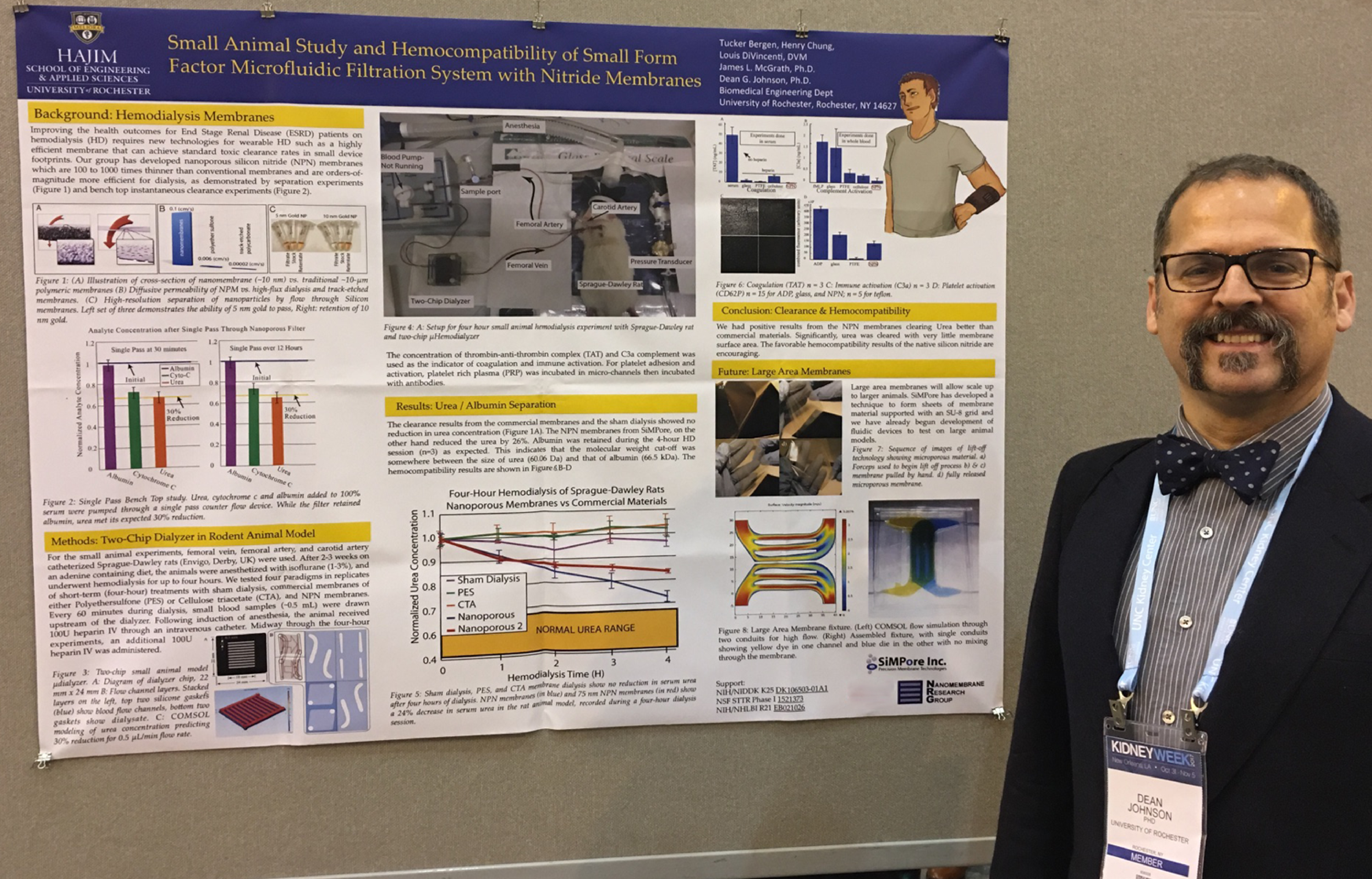
5. Finally the last objective was to learn more about nephrology, renal pathology, hemodialysis, etc by attending presentations and talking with poster authors. Below are some of the presentations and poster abstracts of interest.
Fluid Management in Hemodialysis: Oral Presentations
Thoughts: What I learned here is that nephrologists are still investigating the metrics and correlation to patient outcomes. Currently measuring body weight and ultrafiltration rate. Fluid management is relevant to patient mortality, but the metrology lags behind.
Intradialytic Hypertension Frequency and Short-Term Clinical Outcomes among Hemodialysis Patients
– Jennifer E. Flythe MD, U. of North Carolina
Intradialytic hypertension (ID-HTN) occurs in 5-20% of hemodialysis treatments. Observational data support an association between ID-HTN and increased long-term mortality. However, the short-term cardiovascular (CV) consequences of recurrent ID-HTN are unknown.
What is the best Technology to assess volume status?
This is a work in progress, as talked about in the first talk above. Bioimpedance and bioreactance will help to refine current methods. (weight and UF rate).
Building a Glomerular Capillary Wall: Oral Presentations
Thoughts:
Cellular based technologies are being developed for a number of applications in renal therapy. 1. cure disease in situ 2. repopulate decellularized cadaveric kidneys. 3. populate synthetic scaffolding 4. kidney on chip 5. basic science (not an application, I know)
Genesis of the Renal Endothelia: Emphasis on the Glomerulus
-Seppo Vainio Ph.D. U. of Oulu
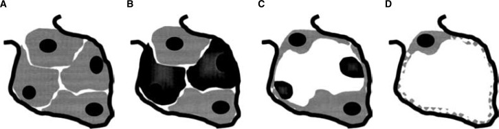
Figure 1. Representation of development of fenestrae. (A) Angioblasts (endothelial cell precursors) invade the capillary cleft as precapillary cords, devoid of capillary lumen. (B) A subset of undifferentiated endothelial cells begins the process of self-destruction (apoptosis). This process is transforming growth factor-β1 (TGF-β1)-dependent. (C) Apoptotic endothelial cells collapse forming apoptotic bodies. Surviving endothelial cells spread to cover the glomerular basement membrane. (D) Endothelial cells extend a thin layer of cytoplasm around the circumference of the glomerular capillary wall. Transendothelial cell pores (fenestrae) develop in this flattened region. Fenestration is vascular endothelial growth factor A (VEGF-A)-dependent.
Renal Carcinoma/Kidney Progenitor Cell Chimera Organoid as a Novel Tumourigenesis Gene Discovery Model
-Seppo Vainio Ph.D. U. of Oulu
Chimeras between embryonic kidney cells and renal carcinoma cells serve as a novel model to assay the roles of co-regulated genes in kidney devel opment and renalcarcinogenesis. They made use of recently developed kidney organ primordia tissue engineering technologies to create novel renal organoids for cancer gene discovery. Then used to examine kidney cancer development
Bioengineering and informatics: Posters
Home Kidney Screening Device
-Ragwa Elsayed MS, San Jose State University
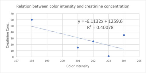
By using the chemical property of picric acid when react with creatinine, a relation is established as a standard curve between creatinine concentration and color intensity.
Using the mobile phone cameras, anyone at home can measure creatinine level by taking an image of test strip.
Parallel Cross-Flow Filtration Microfluidic Device for Renal Micro-Environment Emulation
-Zach Odeh, The First Affiliated Hospital of Dalian Medical University, China
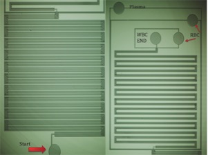
Development of the microfluidic device utilizes photolithography and micro-molding techniques that involve the following basic components: silicon wafer (1 1 1), chrome masks, glass, and polydimethysiloxane (PDMS). Design assumptions called for human blood as the testing fluid. The design utilized a five-row filtration system in the first modular section with varying gap sizes (between pillar edges) from 6.5 microns to 2.5microns. The second modular design included three channels with the main channel flanked by weir micro filtration barriers of consistent gap slits of 3.5 micons.
Between each channel are rows of pillars with spacing designed to filter based on size. Whole blood enters the 5-channel filter and is separated by size, WBCs stay in the center channel, RBCs move to the two channels flanking the central channel, and only plasma moves to the outer channels. The separation is not completed by the end of the 5-channel section, the inner channel (containing WBC and some RBC) continues, splitting into three channels and further separating the blood cells. The outer two pairs of channels simply branch off to further separate the RBC and Plasma. This isn’t exactly what the glomerulus does, and the blood spends a lot of time in the channels.
This step-wise approach, using varying pillar distance is similar to our idea of separating blood components with multiple membranes of different pore size.
A more interesting Glomerulus on a chip (modeling hypertensive Nephropathy) is this paper in Nature.
3D Kidney-on-Chip Platform for Quantitative Screening of Podocyte Structural Integrity
-Evren Azeloglu, Icahn School of Medicine at Mount Sinai
This was simply well structures with some unspecified topography on the bottom of the wells. The authors were not available at the poster (I was busy at my own poster most of the time) They do claim that their 3D wells keep podocytes from dedifferentiating.
Transcriptomic expression of differentiation markers was quantified using RT-PCR. Spatial localization and protein expression were quantified using immunofluorescence. Spatial biomechanics were quantified using atomic force microscope elastography.
Podocytes in 3-D biochips displayed significant upregulation of a wide range of genes associated with the differentiated phenotype. The peripheral processes were selectively enriched for slit diaphragm components nephrin, podocin and neph1 as well as crosslinked actin bundles. Micropatterned podocytes exhibited heterogeneous biomechanical properties with significantly increased elastic modulus in peripheral processes. This spatial phenotype was lost when cells were treated with known nephrotoxic drugs or inhibitors of cytoskeletal integrity. When we looked at phenotypic signatures of protein localization, focal adhesion maturation and cytoskeletal integrity, podocytes in biochips showed reduced cell-to-cell variability and high reproducibility compared to those on unpatterned glass surfaces.
Creating a Roadmap for Innovative Next Generation Renal Replacement Therapy: Breaking the Mould: Oral Presentation
Joe Bonventre, Brigham and Women’s Hospital
There is a good paper on this with a lot of global data.
This was really a presentation of the work the Kidney Health Initiative (of which I have been a part) has done to define and specify the types of devices (wearable, implantable, mechanical, cellular) and the technologies (membranes, cell-lines, bio-reactors) needed to improve patient outcomes.
A new Kidney to Call My Own: Oral Presentations
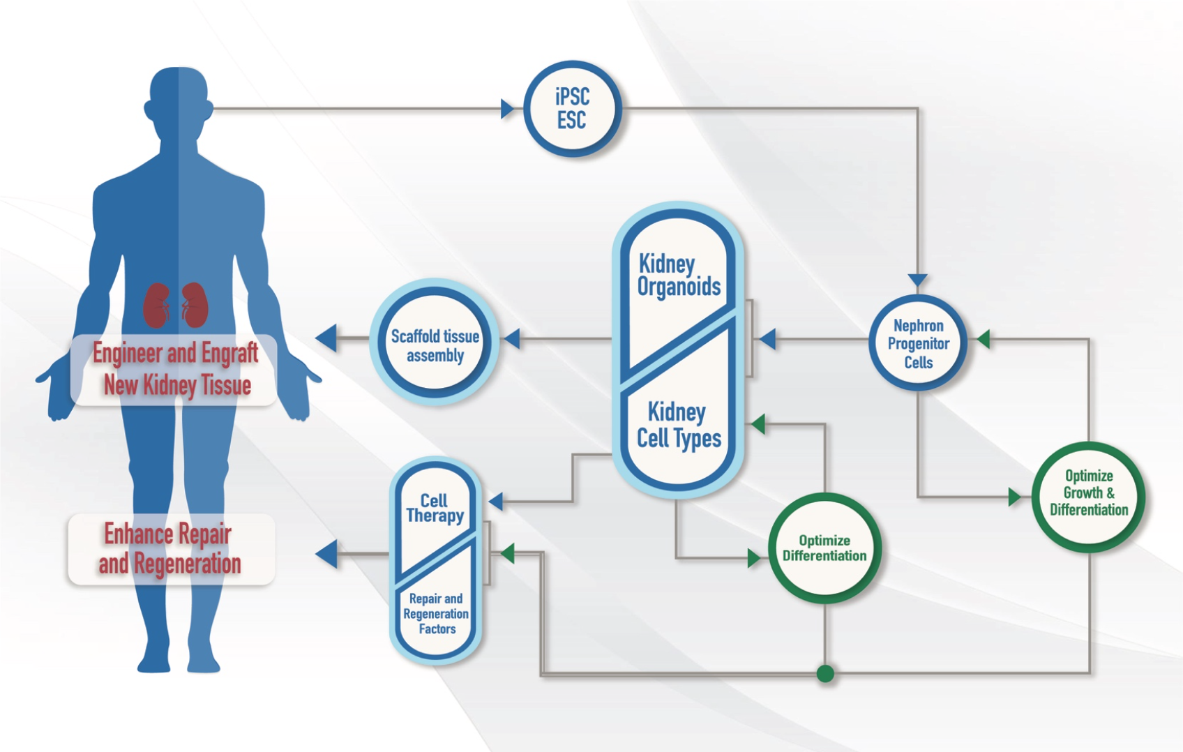
Most of the talks in this session, if not all, were from collaborators in the (Re)Building a Kidney consortium of research projects (RBK). The following image represents the goals of RBK. If I need collaborators in the future, this would be a place to look. It also give a good cross section of the research landscape. (Can you cross section a landscape?)