Size Exclusion of the Vasculature – Polarized TNF-alpha Revision Experiment
Background
Previously we characterized HUVEC monolayer barrier via TEER in traditional transwell systems following polarized TNF-alpha exposure (Figure 1). One major shortcoming of this experimental set up for our study is it does not accurately reflect the permeability of HUVECs within our uSiM system (where all other experiments were performed). Thus, a couple reviewers wanted us to measure small molecule (dextran) permeability within our uSiM system specifically. On top of this, they wanted to ensure the results were consistent across two dextran sizes: 10 and 70 kDa.

Methods (updated for revision)
Small Molecule Permeability
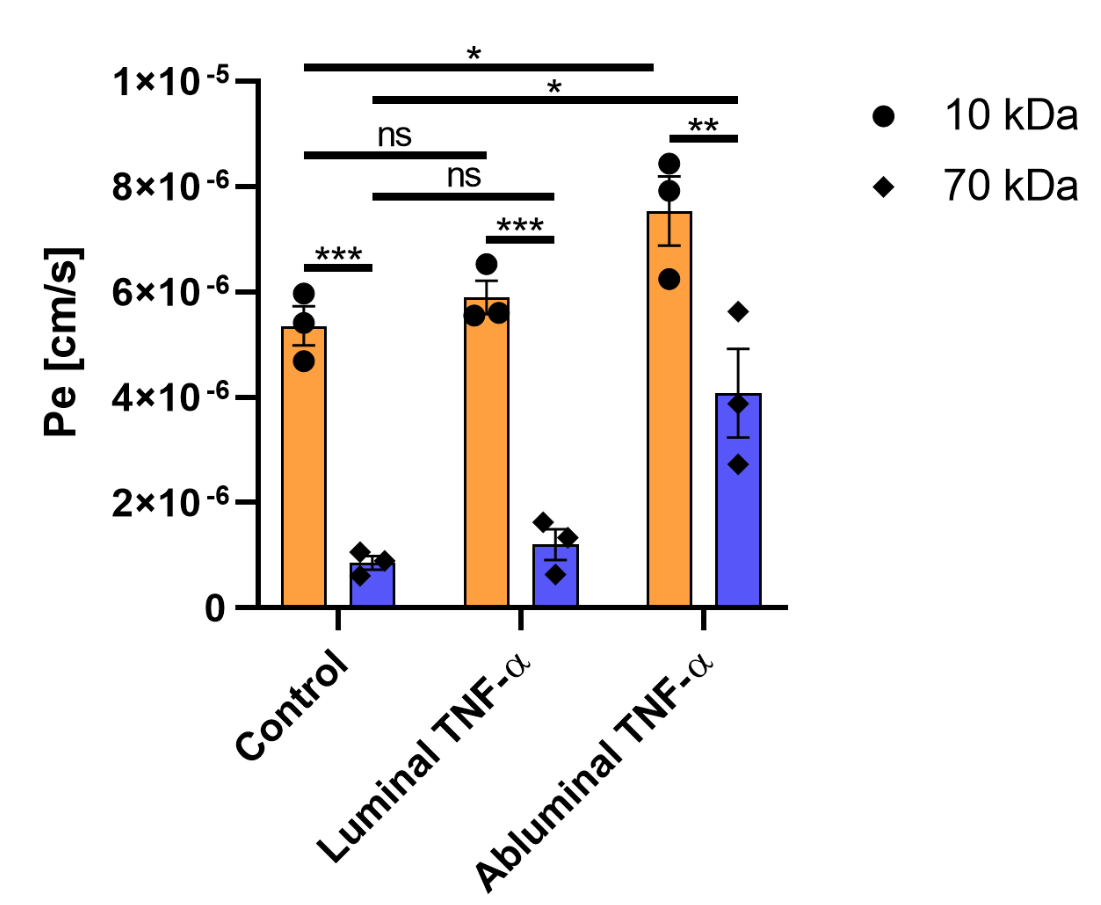
HUVECs were seeded in μSiM-MVMs as stated previously. Once confluent (24 h post-seed), HUVECs were exposed to control medial or TNF-α [20 ng/mL] media (luminally or abluminally) for an additional 24 h. Following treatment, devices were loaded luminally with 10 or 70 kDa FITC-dextran [1 mg/mL] (Sigma, St. Louis, MO) in MCDB-131 complete media and returned to the incubator (37 °C, 5% CO2) for 1 h. Media was gently sampled from the abluminal channel and transferred to a black 96-well plate. Sample volumes were adjusted to 50 µL to allow for accurate readings. Depending on the tracer used, a 10 or 70 kDa FITC-dextran reference ladder was prepared as follows: 200, 100, 50, 25, 12.5, 6.25, 3.125, 0 μg/mL in MCDB-131 complete media. Reference samples were transferred in 50 μL aliquots to the sample 96-well plate. Fluorescence intensity was measured at 490/520 nm Ex/Em in a microplate reader (Tecan, Männedorf, Switzerland). Permeability coefficients (Pe) were calculated by inputting the FITC-dextran flux into equation (1):
Pe = Jf/ACi (1)
where Jf is the flux of FITC-dextran into the abluminal channel (mass/time), A is the membrane area (0.014 cm2), and Ci is the concentration of FITC-dextran added to the luminal channel (1 mg/mL).
Results

Conclusion
The uSiM-MVM has characteristic size exclusion properties of a native vascular barrier. Interestingly, abluminal TNF-alpha significantly increases small molecule permeability disproportionately compared to luminal TNF-alpha. The degree of disruption appears to be significant based on Pe/D analysis.