HMM vs. LMM diffusion
My last post dealing with this project was the first to definitively demonstrate HMM diffusion through pnc-Si. Unfortunately, there wasn’t a difference between the HMM filtrate signal for intact and digested (+RNAse) samples. As a reminder, HMM is a large protein multimer held together, partly, with RNA. We hope to show that HMM can not diffuse through pnc-Si pores because of its large size but if you digest HMM into lower molecular weight (LMM) components, those LMM pieces can diffuse through. Something that breaks up HMM could be considered a ‘hit’ in a high throughput screening assay for HIV drugs.
I wondered if both samples were broken down in the last experiment so that the same amount of protein was able to pass. So, I got a batch of lower molecular mass A3G (LMM) to test alongside high molecular mass A3G (HMM). I set up a diffusion experiment in SC 364 square Sepcons (same wafer as last experiment) with 22uL of 2.2mg/mL protein in the Sepcon and 22uL HMM buffer in the closed bucket. Samples were centrifuged at 13000 rpm for 10 minutes before use. I let these samples run for 24 hours at 4C and then used the higher sensitivity Quant-IT assay to quantify signals.
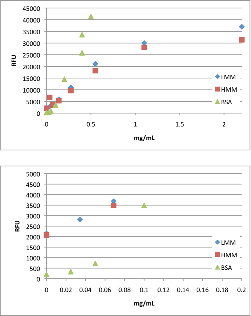
Here are the full (top) and low concentrations (bottom) standard curves of HMM, LMM and BSA. One of the claims of the Quant-IT assay is that there is little variability in signals between proteins, so the drastic difference between A3G and BSA is disconcerting. There are a couple possible reasons for this discrepancy: 1. the RNA in HMM/LMM is interfering with the Quant-IT reagent 2. the protein concentration of HMM/LMM is slightly lower than what I report because I don’t know the 280/260 ratio (the relative amount of protein and RNA). Fortunately, the HMM standard curve is linear up to ~ 1mg/mL.
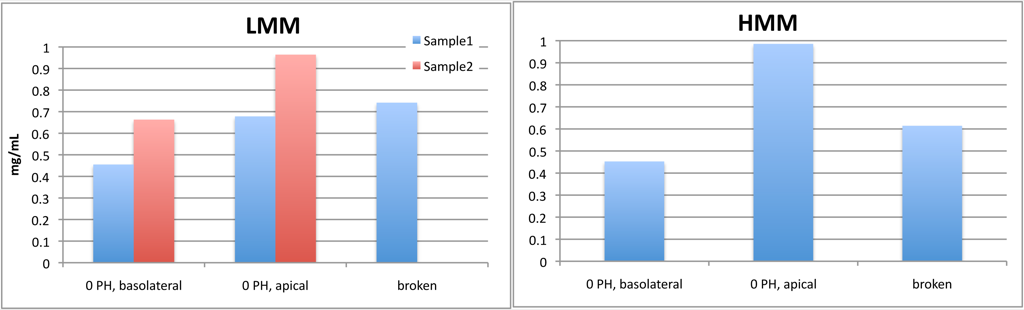
Here are the experimental samples. I included broken samples as positive controls (which should have mixed entirely to give a 1.1 mg/mL equilibrium concentration of protein. These broken samples gave concentrations < 1.1 mg/mL which indicates that there is loss of protein in this set-up (adsorption to Sepcon plastic, pnc-Si and/or PDMS). For both HMM and LMM, the apical well has a higher concentration than the basolateral well, which makes sense. However, the concentration is less than the expected equilibrium concentration which further suggests protein loss. Both HMM and LMM diffused across SC 364 since there protein was detected in the basolateral volume. However, it doesn’t seem like the LMM concentration is significantly higher than the HMM concentration, which was the expectation. So, it was encouraging to detect HMM and LMM in the filtrate (again) but I can’t say that there was a difference in diffusion between low and high molecular weight samples.