Introduction of beads into the flow module
1. Introduction
Fluid flow is one of the key elements of vascular models for delivering nutrients and growth factors to cells as well as introducing other components like neutrophils. In post 1, we described our modular design for incorporating a flow module into the uSiM device. Then, we presented fluid flow simulation and its experimental validation in post 2. Here, mathematical calculations for the deposition of beads and neutrophils on the membrane are reported. Then, pulling fluorescent beads experimentally and depositing them on the membrane are demonstrated.
Table 1. Milestone of R61 project – Abhyankar lab
|
Tasks |
Status |
| Flow module design | Presented in Post 1 |
| Flow models | Presented in Post 2 |
| Introduction of beads/neutrophils | Presented here |
| Flow module testing | In progress |
| TEER module | Future work |
| Side-by-side multi-cell culture | Future work |
2. Method
Experiment setup: All tubing, connectors, and reservoirs were primed with DI water. A mixture of 5um Fluorescent Polystyrene Latex particles (2.5% w/v) (Magsphere, CA, USA) with DI water was created at a ratio of 1 to 99. Then, the mixture was pulled out from the reservoir into the device using a syringe pump at a flow rate of 10 ul/min (Figure. 1).
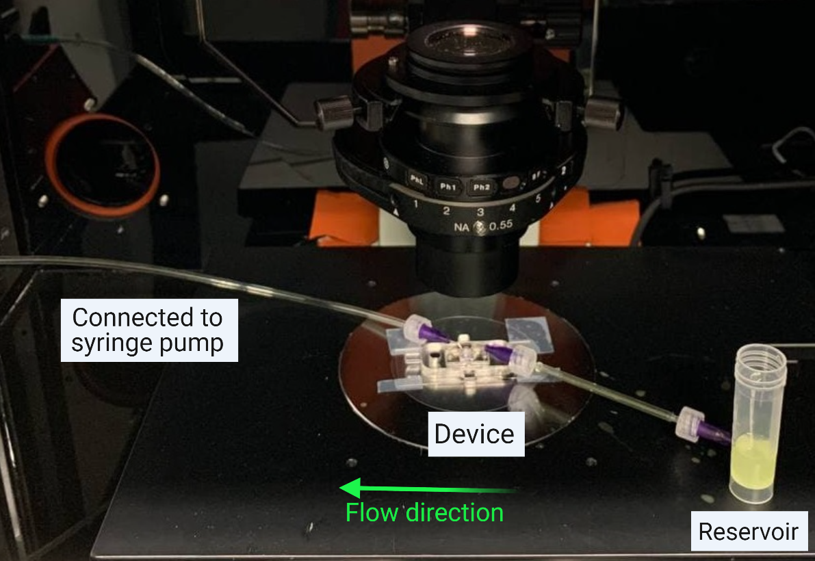
Figure 1. Experimental setup for pulling out beads/neutrophils.
Assumptions:
- The fluid is incompressible
- Fully developed flow
- Particles follow the fluid velocity
- Particles are spherical and rigid
- Particles do not affect the flow and do not interact with each other (dilute)
Stokes’ law:
As a particle moves through the microchannel along with the flow, it spends a certain amount of time over the membrane (residence time, tf = L/Vf, where L is the length of the membrane and vf is the velocity of the fluid). Because of the parabolic velocity profile, the fluid velocity is a function of the position above the membrane (zero at the surface, and a maximum at the midpoint of the channel). Assuming the velocity of the particle matches the fluid velocity (dilute bead solution with low Reynolds number) there is another time scale (ts = h/vs) that describes the time it takes for the particle to settle from a position h onto the membrane surface where Vs is the settling velocity (see Figure 2).
In a situation where the settling time is less than the residence time (ts/tf < 1), the particle can settle onto the membrane before it is swept past the membrane. The settling velocity can be calculated using Stokes’ Law:

Figure 2. Settling velocity and settling time over distance h for a particle based on Stokes’ law
3. Results
Settling velocity and height threshold: Considering DI water as the medium with a density of 993.3 Kg/m^3 and viscosity of 6.9*10^-4 Pa.s at 37 C (Ref), the settling velocity of polystyrene beads and neutrophils is calculated for a flow rate Q = 10 ul/min (to ensure there is no damage to cells based on previous simulations – reference NRG post 2).
The height h at which ts / tf = 1 is calculated for a polystyrene bead and a neutrophil assuming a given diameter, d is shown in Table 2. We have previously validated our flow and COMSOL simulations, and here we use Vf from our COMSOL model and calculate Vs using Stokes Law. Thus, beads at a position h<12 um and neutrophils at a position of h<36 um, could settle onto the membrane. If we assume that beads/neutrophils are distributed uniformly in the mixture, we can expect that 6 % of beads and 18 % of neutrophils could settle on the membrane at a flow rate of 10 ul/min.
Table 2. Height threshold of polystyrene beads (p=1055 Kg/m^3) and neutrophils (p=1080 Kg/m^3) at Q= 10 ul/min.
| Particle type | d (um) | Height threshold (um) | particles that could settle onto the membrane (%) |
| Polystyrene beads | 5 | 12 | 6 |
| Neutrophils | 12 | 36 | 18 |
Bead settling: Click here to watch the captured video during the experiment at a flow rate of 10 ul/min.
4. Conclusion
The preliminary results presented here can be used to estimate starting concentrations and flow rates that promote neutrophils settling onto the membrane. The number of neutrophils on the membrane can be increased by decreasing the flow rate, increasing the concentration, or increasing the time the flow is maintained (introduction time).