iPSC-derived Brain Pericyte-like Cells
Introduction
Pericytes are mural cells that line the outer surface of microvessels and regulate diverse aspects of vascular development and function. We differentiate brain-pericyte-like cells (BPLCs) from hiPSC clones using the protocol published by Eric Shusta’s lab (Gastfriend et al., 2021) (Figure 1). This protocol describes methods to differentiate hiPSCs to BPLCs via neural crest intermediate, that mimics the developmental origin of forebrain pericytes.
We first direct hiPSCs to neural crest stem cell (NCSC) differentiation over a 15-day long period. We isolate p75-NGFR+ NCSCs via magnetic-activated cell sorting (MACS) on day 15 and then replate them for pericyte differentiation. After approximately 7 days, we obtain BPLCs that are defined with the NG2+PDGFRβ+αSMAlow pattern.
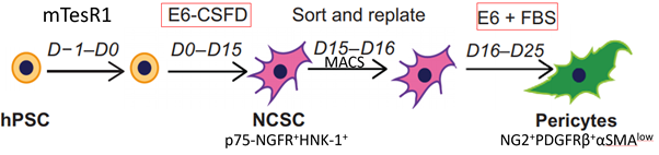
Figure 1: Schematic representation of the brain pericyte-like cell differentiation protocol.
Results
Phenotypic characterization of hiPSC-derived brain pericyte like cells
We validated NG2+PDGFRβ+αSMAlow brain pericyte-like cells using flow cytometry staining and immunocytochemistry (Figure 2).
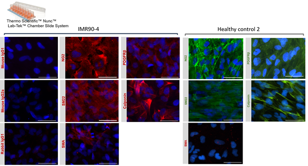
Figure 2: Phenotypic characterization of hiPSC-derived NG2+PDGFRβ+αSMAlow BPLCs. Immunofluorescence staining of pericyte markers PDGFRβ and NG2, mural cell-associated proteins SM22 and calponin and α-smooth muscle actin (SMA) on day 25 ( IMR90-derived BPLCs) and on day 26 (HC2-derived BPLCs). Cell nuclei were counterstained with DAPI. Scale bar is 50 μm.
EECM-BMEC-like cell and BPLC co-culture
The BPLCs and EECM-BMEC-like cells were fluorescently labeled using cell tracker green and red, respectively. The BPLCs were seeded into the bottom channel and the devices were incubated in an upside-down position so that BPLCs attach to right underneath the membrane. After BPLCs attached, the chips were turned back to the upright position and the EECM-BMEC-like cells were seeded into the well. We showed that BPLCs remained attached to the upper walls of the bottom channel and maintained contact with EECM-BMEC-like cells through the nanopores of the membrane.
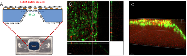
Figure 3: Co-culture of EECM-BMEC-like cells and BPLCs in μSiM device. (A) Graphical representation of the position of EEC-BMEC-like cells and the BPLCs. (B-C) The BPLCs (green) formed a monolayer on the upper wall of the bottom channel and along the trench. (B) top view (C) side view.