Fibroblast Wound Healing with EVs and EV Depleted Media
Introduction
Previous experiments completed by Hayley Miller investigated the effect of small extracellular vesicles (EVs) collected from Adipose Derived Stem Cells (ADSCs) on wound closure velocity and cell speed. Her previous related post can be found here. It was initially hypothesized that EVs play an important role in wound healing and affect the wound closure velocity. Results from her experiments, summarized in Figure 1a and 1b, rejected this hypothesis and indicate that EVs do not have as significant an impact as originally thought. Based on literature read by our lab, we believe past positive EV results from others are a result of co-precipitation of growth factors during EV isolation. We now hypothesize growth factors have a larger positive impact than EVs on wound closure velocity. We tested this hypothesis by adding EV depleted media to our wound healing experiments. We believe our EV isolation process effectively separates growth factors from EVs and our added EV depleted media condition supports this belief (see Methods and Results). Since EV depleted media contains growth factors, it was expected that cells healing in EV depleted media would heal faster than cells healing in a small EV solution.
Figure 1a and 1b: Summary of Past Results
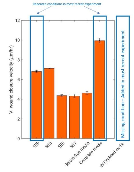
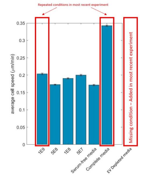
Past wound closure velocity and average cell speed results outlining which conditions were repeated and which were added.
Methods and Results
ADSC cells were grown in complete media until approximately 80% confluent. Once confluent, the complete media was switched with serum-free media for 24 hours. The media from the flasks was collected and small EVs were collected after the fourth spin, two hours at 100,000 g, in the EV isolation process.
Human Dermal Fibroblasts (hDFbs) were seeded in a 24 well plate and grew until 90% confluent. A wound was created in each well by scratching down the center of the well using a p200 pipette tip. Cells were left to heal in complete media, EV-depleted media, or serum-free media supplemented with isolated EVs (1E+9 EVs/mL) for 48 hours. Time lapse videos for all conditions were recorded and an example of wound closure over 48 hours can be seen in the link. Complete media acted as a positive control and also allowed this experiment to be accurately compared to Hayley’s past experiments. Results for the time lapse were analyzed by tracking individual cells using the MATLAB tracking code created by Henry.
Nanoparticle Tracking Analysis (NTA) was completed on the serum-free media supplemented with small EVs to confirm the EVs were successfully isolated. Analysis done on the small EV solution confirmed the presence of small EV sized particles at a concentration of 1.7E+9 particles/mL. Analysis done on the EV depleted media confirmed a lack of particles in the small EV size range throughout the media.
The results for this experiment were as expected. As summarized in Figure 2a and 2b, the complete media had the fastest wound closure velocity and average cell speed. The cells healing in EV depleted media had a faster wound closure velocity and average cell speed than the cells healing in the small EV solution. This confirms that EVs are not as influential in promoting wound healing as previously expected. Shown in Figure 3, the complete media and small EV (1E+9) results and trends were comparable to Hayley’s previous results which allows us to believe the cells are behaving similarly across all experiments.
Figures 2a and 2b: Summary of Most Recent Data
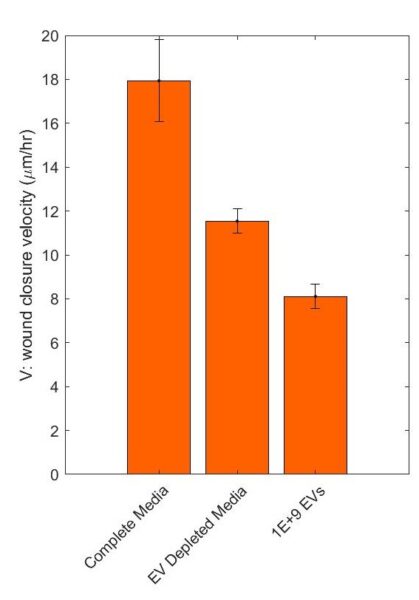
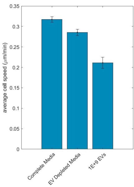
Wound closure velocity and average cell speed for complete media, serum-free media, and serum-free media supplemented with small EVs.
Figure 3: Comparison to Past Results
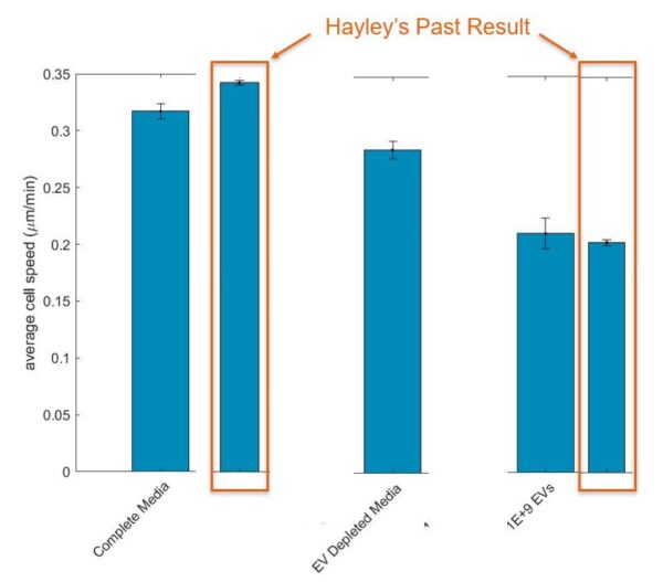
Current results compared to past results for repeated conditions.
Next Steps
I plan to complete one final wound healing experiment investigating four conditions: complete media, serum-free media, EV depleted media, and serum-free media supplemented with a higher concentration of EVs (5E+9 EVs/mL). The complete media is the positive control. The serum-free media is the negative control. The hypothesis that growth factors have a greater positive impact on wound closure velocity will be confirmed if the cells in EV depleted media heal significantly faster than the cells in the high concentration of small EVs.
The EV depleted media and serum-free media supplemented with small EVs will also be analyzed using an ELISA kit to confirm the presence, or lack thereof, of growth factors in the EV depleted media and small EV solution. VEGF and PDGF will specifically be investigated.