Flow Based HUVEC Alignment – Preliminary Data
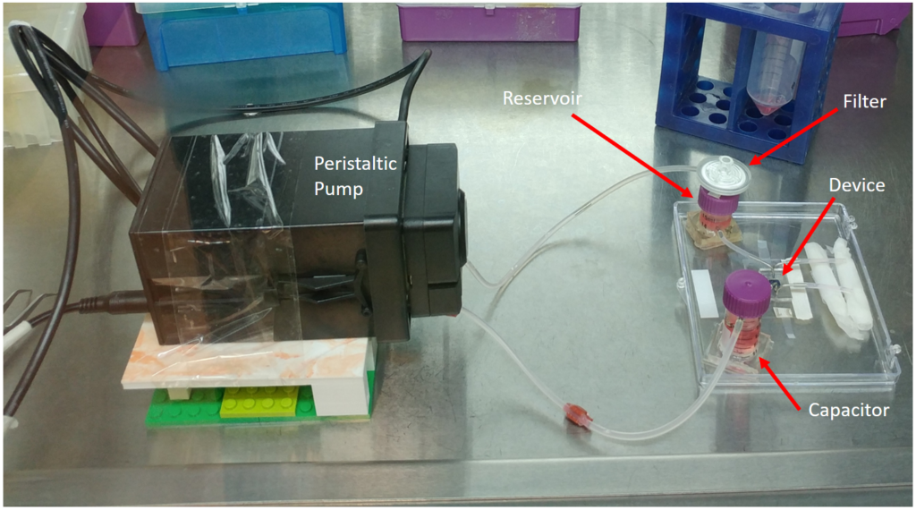
As a follow up to my previous post, I would like to present some preliminary data on the subject of endothelial cell alignment under shear stress with variable substrate stiffness. The flow set-up (Figure 1) allows for steady flow across the membrane on which the HUVECs are cultured.
No Gel HUVEC Alignment
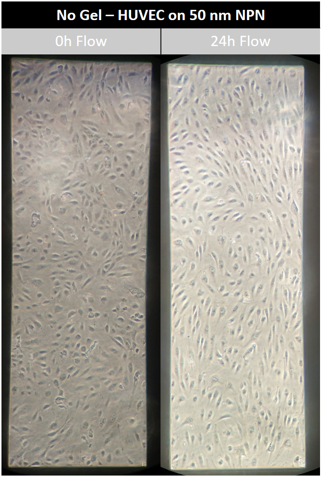
Alignment of HUVECs on the bare NPN membrane was previously observed. In order to see how reproducible this observation was, the experiment was repeated under the same conditions. The results show alignment after 24 h flow (Figure 2, not quantified). This outcome justified pushing forward to the next experiment, which is observing the effects of a gel on the underside of the NPN chip with respect to HUVEC alignment.
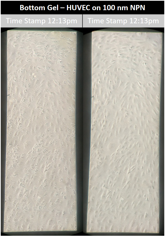
Underside Gel HUVEC Alignment
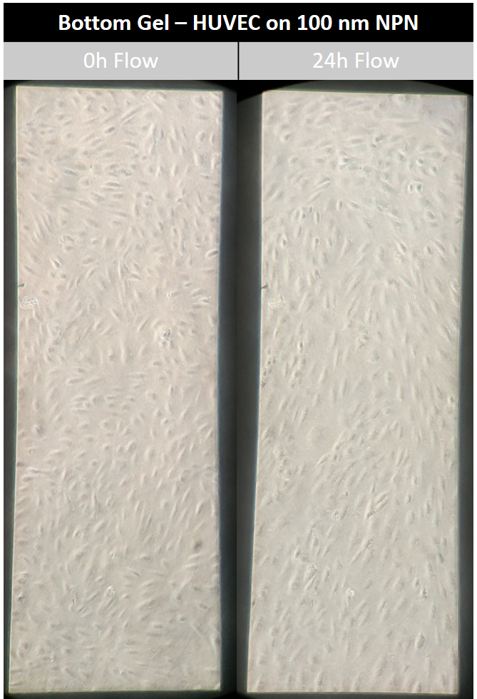
Given previous data (see previous post), we hypothesized that endothelial cells cannot align properly on the gel covered NPN membranes due to mechanical changes in the substrate on which the cells are being cultured. In order to rule out the possibility that the underside gel is chemically altering the ability of the endothelial cells to align, we needed to perform a reasonable control. Type I Rat Tail Collagen gel was added to the bottom surface of 50 and 100 nm NPN chips and HUVECs were subsequently culture on the top side (non-gel containing side). The flow set-up was assembled as previously described and the HUVECs were subject to 10 dyn/cm^2 flow for 24 h. Results on both NPN thicknesses showed endothelial alignment following 24 h flow (Figures 3 & 4).

HUVEC Vacuole Formation