The Concentration of the Barcikowski Peptide is at the Lower Bound of Our Detection Limit
Recall that we’ve been having a great deal of trouble following the free ligand during our separations of the Bioconjugated Barcikowski particles, and that this trouble also means we can’t see the ligand after centrifuging the pure sample. In the interest of thoroughness, I replicated the assay the Barcikowski lab used to quantify the unbound ligand in their recent paper(Penetratin-Conjugated Gold Nanoparticles). Note that they are using a ligand called penetratin in this paper, which is not what they have sent us – we are working with a peptide that has a strong negative charge and the sequence CEEEEEEEEEEEEW. Nevertheless, we thought following their protocols exactly was worth doing. There, they spun the conjugated gold down for 30min at 40,000g and used UV-Vis spectrometry (Shimadzu 1650 – tech specs) to quantify how much ligand remained suspended. Repeating this, we got the following curves:
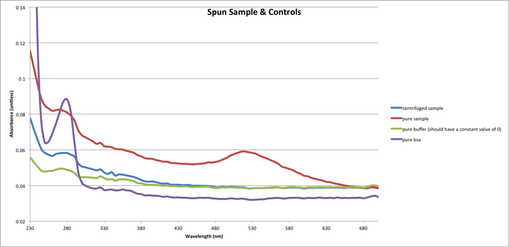
Which looks great, except for that weird buffer curve. In the interest of fixing the oddball measurements we sometimes get from the TECAN, I emailed the company and described our phantom 280nm peaks. I hoped they might have some secret technique for cleaning the plate more thoroughly. They asked me for a totally blank plate:
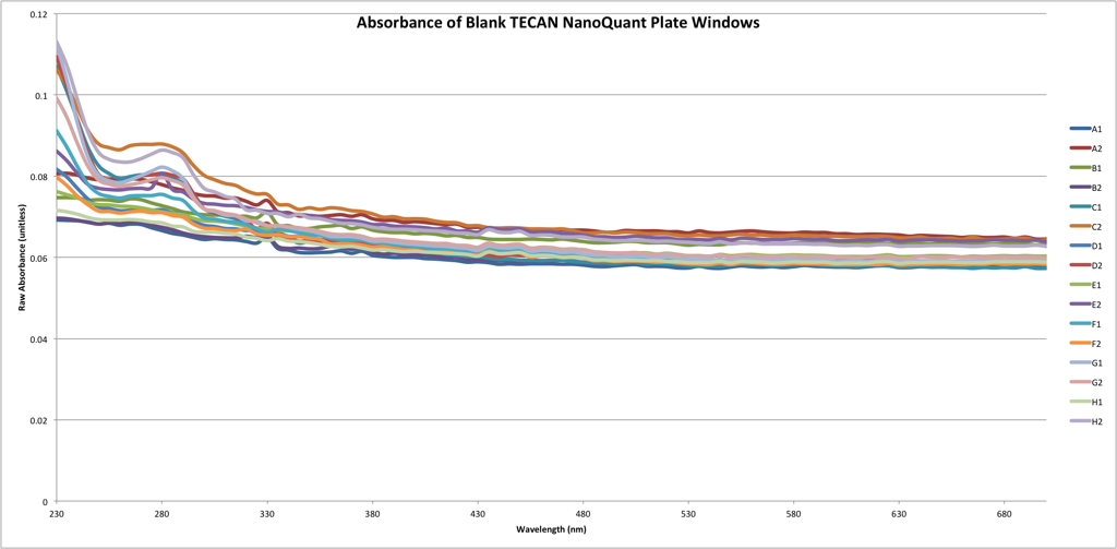
The persistance of that peak at 280 in some of the windows, and the fact that none of the measurements come close to perfectly overlapping, is depressing. Hopeful TECAN will help us out.
Next we wondered if we were simply running up against the detection limits of the TECAN generally, and if it might be better to either switch to the other spectrometer in the basement (a BioRad SmartSpec 3000) or purchase the Nanophotometer and use it for detection. First, we found the molar concentration of free ligand:
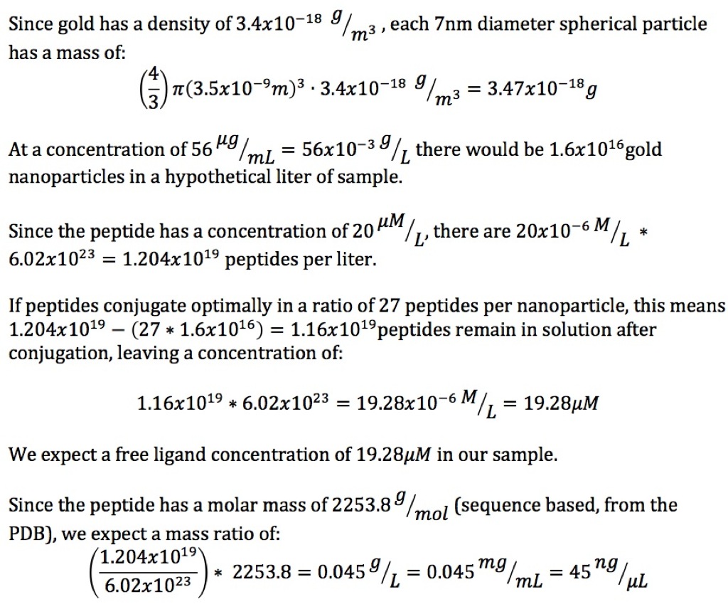
Giving us a protein concentration of 45ng/uL, or 0.045mg/mL. Note that the act of conjugation only reduces the concentration of the peptide by about 4%. The Nanophotometer P-Class (tech specs) claims a BSA detection limit of 0.08mg/mL and a DNA detection limit of 2ng/uL – both in the small volume setup (with larger volumes that lower bound becomes BSA 0.02mg/mL and DNA 0.5ng/uL). Our other Spectrometer – the BioRad SmartSpec 3000 (tech specs – look at pg. 49) doesn’t list it’s detection limit, but this picture: 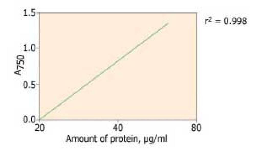
makes me think the lower bound is 20ug/mL = 20ng/uL. Surprisingly, it’s the TECAN m200 (tech specs), which is what we’ve been using, that wins the lower detection limit game, with an unlisted BSA detection limit but a DNA limit of 1ng/uL. I believe our problem is with the plate, not the machine.
Another avenue of attack is getting our hands on free peptide and making our own standard curve. With free peptide we could also do a model experiment and separate out gold nanoparticles we add from a higher concentration of peptide. To this end I’ve asked the Barcikowski lab to ship us some solutions of the peptide they are using.