HUVEC Growth on Si
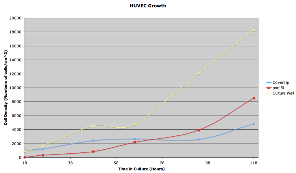
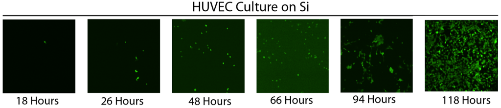
Over the past five days Anant and I monitored human umbilical vein endothelial cell (HUVEC) proliferation on three substrates: RTP-treated Si samples (both with and without membranes), glass (microscope coverslips), and plastic (6-well tissue culture plates). After the first two time points very few cells were observed on the silicon samples, so we added more cells to all samples. Images were taken at 10x magnification using the live/dead assay to visualize the cells.
Interestingly, membrane samples at time points 96 and 118 hours were discolored, whereas Si samples with no membranes (solid Si) did not discolor.
It is clear that HUVEC proliferate and form monolayers on Si. We plan on repeating this study again for both HUVEC and fibroblasts, with the addition of monitoring membrane discoloration.
For some reason you have to right click on the first image to open it, even though they are both the same file type….
That’s odd… I fixed the link by clicking on the picture and manually inserting the address in the editor.