HUVEC spreading on various microporous SiO2 membranes
This is part of our work studying how cells adhere, spread and proliferate on varying pore size/porosity SiO2 membranes. This post will be a qualitative look at how HUVEC spread over membranes by staining for actin filaments. In a future post we will quantify the degree of spreading at 1hr, 4hrs and 24 hrs.
Calculation of porosity
These membranes are lithographically patterned on a hexagonal array such that the center-to-center pore spacing is either 2 or 4 pore diameters.
Low Porosity = 5.7%
High Porosity = 22.7%
Porosity-calculation
These are the membranes that were tested and at the current time represent all available lithography masks (pore size in microns):
0.5 LP, 0.5 HP, 3.0 LP, 3.0 HP, 5.0 LP, 8.0 LP
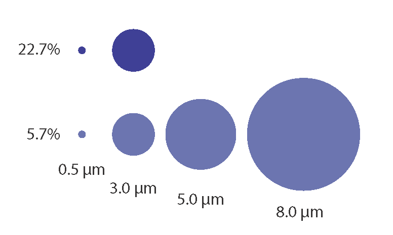
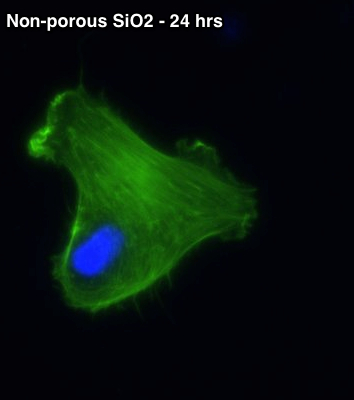
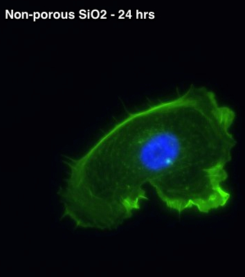
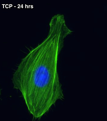
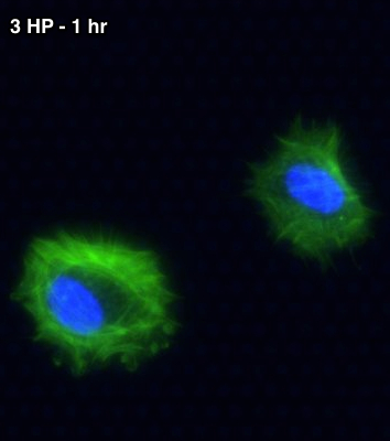
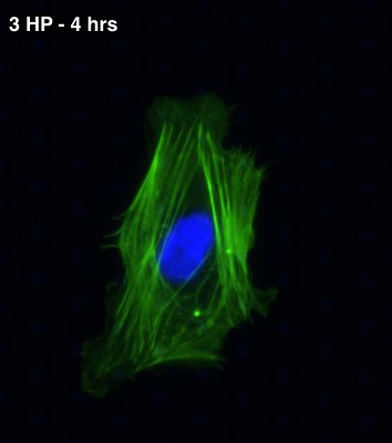
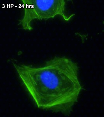
Membranes were prepared by treating with 1% Geltrex in PBS for 30 minutes at room temperature. Solution was aspirated off and surfaces were allowed to dry prior to seeding cells. The chamber below the membrane was filled with 30 uL of M200 media. HUVEC were passage 5 and seeded at approximately 300 cells per window (20 uL). For the 1 hr time point experiments, the samples were washed with PBS, then fixed, permeabilized and stained with DAPI (blue) and phalloidin (green). For all other experiments (4 hr and 24 hr), wells were flooded with 800 uL of warmed M200 media at 1 hr and returned the incubator until they were washed, fixed and stained at either 4 or 24 hrs. A minimum of 2 membrane windows were used at each time point and condition. Geltrex pre-treated non-porous SiO2 membranes and tissue culture plastic (TCP) were also used.
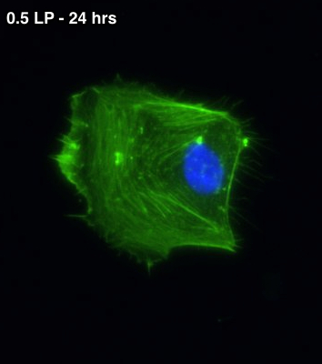
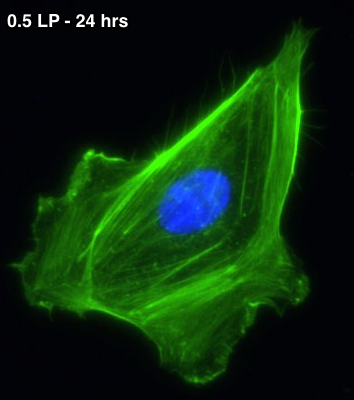
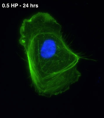
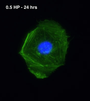
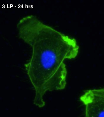
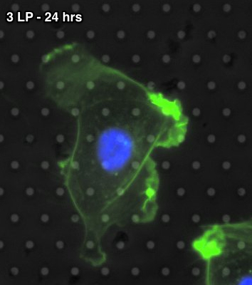
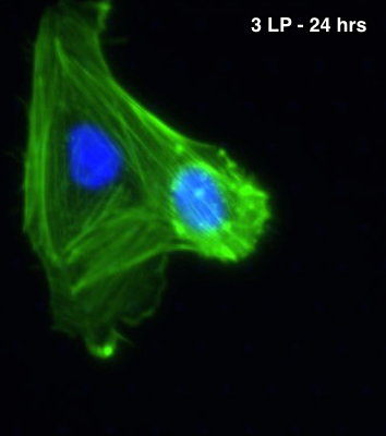
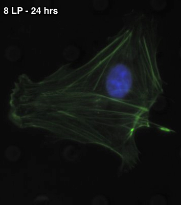
It appears that HUVEC attempt to “poke” or “peek” through membranes with 3 micron pores. The degree of poking is greatest on the 3HP membranes, but is also noticeable on the 3LP. There may be some cell protrusion through the 5LP membranes, but it is minimal compared to the 3LP and 3 HP membranes. Surprisingly, the cells appear to glide over the pores in the 5LP and 8LP membranes. Images are also shown of cells spreading at 1 hr and 4 hr on the 3HP membranes at end of the page. There are no obvious protrusions through the membrane at these early time points. Qualitatively, it appears that up to 1/3 of cells on the 3LP and 1/2 of cells on 3HP membranes have obvious protrusions through the membrane.
All of the following images are 100 microns wide (40x LWD objective):
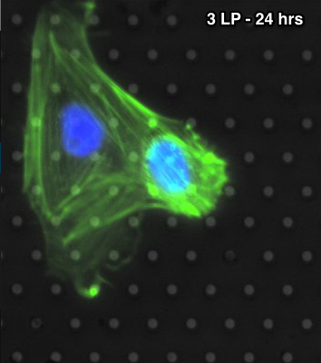
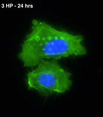
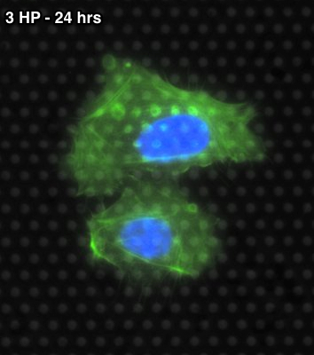
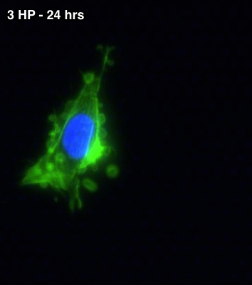
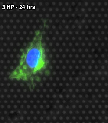
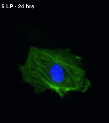
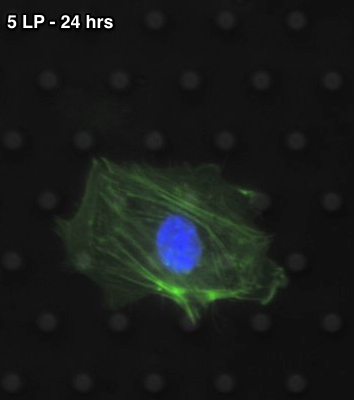
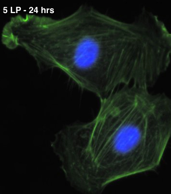
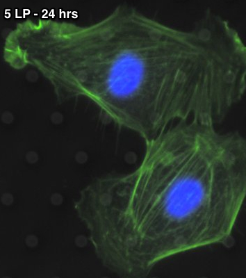
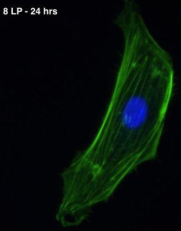
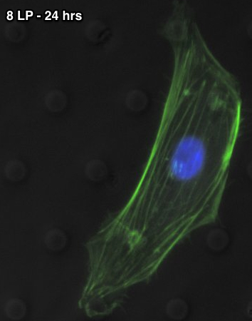
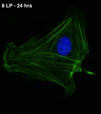
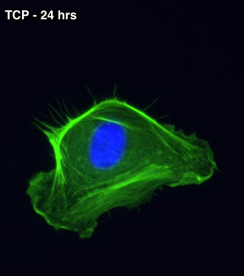
The following are independent 1, 4 and 24 hr spreading pictures of HUVEC on 3HP SiO2.
Which media is this-M200? Is it proprietary or something you prepare in-house?
Also, how is this spreading in comparison to that on 3 and 8 micron PC/PTFE membranes present in the transwells?
M200 is a proprietary EC media from Life Technologies.
We have not looked at spreading at early time points on TE membranes. Background fluorescence signal after staining can sometimes be problematic and the randomness of pore locations as well as doublets could complicate interpretations.