Separation of Microbeads from Whole Blood on 8 and 9 Micron Slit Membranes
A few weeks ago, Josh and JP brought me some 3 window microslit membranes that had edge aligned 8 and 9 μm slits. While these may seem to be quite large for most applications, they are in fact perfect for working with whole blood. The experiments that we have planned for these membranes are based on the ability to separate out certain particles from blood. Specifically, the goal is to be able to spike in microbeads that have been coated with an antibody specific to antigens on certain biomarkers of interest (e.g. circulating tumor cells) and then separate out these beads based on their size in a SepCon format.
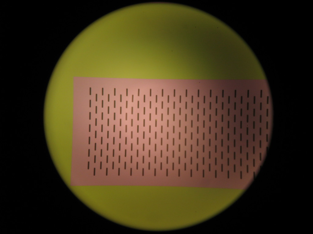
Figure 1: New 3 window membranes with 8μm slits that have been edge aligned. Burst pressures are around 30 psi because of the edge alignment.
To initially test the viability of these membranes for separating beads from solution, I found some 10 μm polystyrene beads and suspended them at a concentration of 1.84 x 10^10 beads/mL in nanopure water. This suspension was very cloudy, almost the color of milk. I then took approximately 200 μL of this suspension and loaded it into a SepCon cup and spun it through for 5 minutes at 2000 rpm. After it was done, the filtrate was clear and the surface of the membrane was white from the beads. There was a small droplet of water on the surface of the membrane, but pretty much everything had passed through. I then took the membrane out of the chip and looked at it under the microscope. Not having payed too much attention to the concentration of the beads, they were stacked everywhere on the membrane and there were many layers of beads, as can be seen in Figure 2.
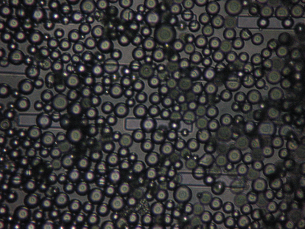
Figure 2: Reflection bright field microscopy image of 8 μm slit membrane surface with 10 μm ploystyrene beads after spinning. There is a noticeable lack of alignment of beads on the slits in this image.
Realizing that this concentration was too high for good image quality and that we wouldn’t be using this high of a concentration of beads in the actual experiments, I diluted the beads 1/100 in nanopure water again to reduce the concentration and hopefully see some alignment on the slits rather than just stacking of the beads. This concentration was still quite high and there was a cake layer beginning to form, but there was also noticeable alignment on the slits. The slits are 50 μm in length, so nominally 5 beads can fit along their length. We can see this effect in Figure 3.
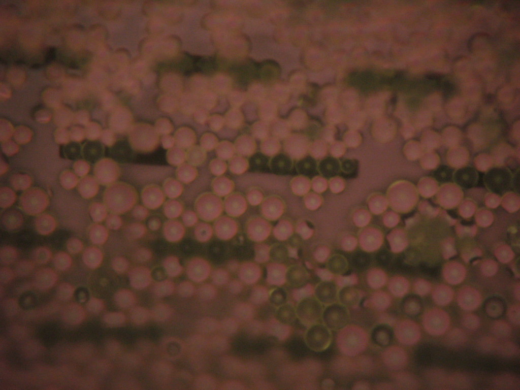
Figure 3: Diluted microbeads on 8 μm slit membranes. The beads are starting to align in the pores, but the water is still able to pass through which indicates that there is still space available for cells to squeeze through.
Since I was seeing alignment, I decided that I could try to run the experiment now with blood and the untreated beads. This simply as a proof of principle experiment that I could pass blood through even with beads in the slits. I took 1 μL of bead solution and added it to a mixture of approximately 50 μL of my blood (from a finger prick) and 100 μL of sodium heparin solution (to prevent clotting). After spinning for 15 minutes (to ensure that the surface of the membrane was dry for imaging) at 2000 rpm, I was quite surprised to see that all the cells had passed through the membrane and were pelleted on the bottom of the tube. Furthermore, as can be seen in Figures 4 and 5, the beads were captured on the slits quite nicely.
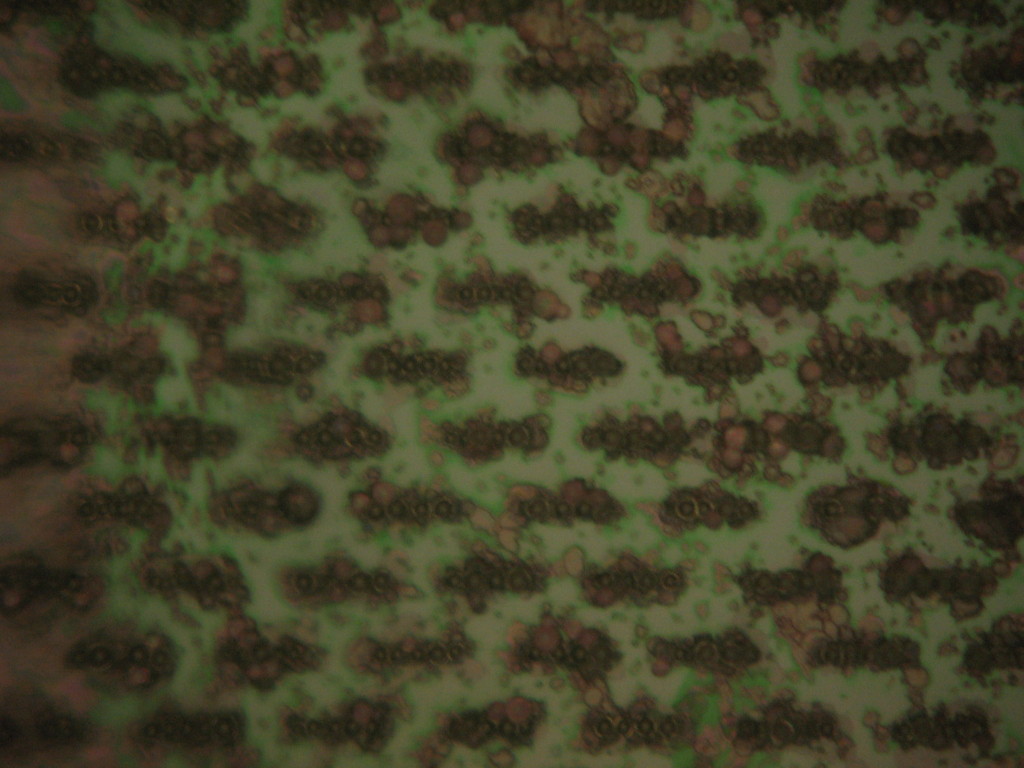
Figure 4: Reflection bright field microscope image of beads aligned in the slits after passing whole blood through. Note that there is some junk on the surface of the membrane and around the beads, but this is likely just lysed cells and debris.
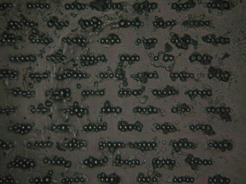
Figure 5: Transmission bright field microscopy image of the beads on the slit membrane. This image shows the alignment of the beads in the slits much better.
With this preliminary data, the next step of the experiment is going to be demonstrating that we can spike a sample with beads coated with antibodies and have them be captured and retained in the slits while the remainder of the sample passes through. The preliminary experiment would be using whole blood and beads coated with anti-CD71 (which is the transferrin receptor) and trying to isolate reticulocytes (immature red blood cells) from the whole blood. If this experiment works, it will demonstrate that we can indeed capture particles of interest and opens the door to a wide range of biomarkers that can be found in any bodily fluid.