Air-Blood Barrier TEER Results
I finished 3 independent experiments of air-blood barrier co-cultures – these consist of bEnd3 endothelial cells on 1 side and RLE-6TN lung epithelial cells on the opposite side of the membranes. We didn’t expect these cell types to potentiate TEER in anyway (like we expected glial cells to tighten endothelial barrier function). However, our collaborators in Environmental Medicine are interested in these type of co-culture models to study nanoparticle translocation from the lung to the blood.
I did 2 experiments with bEnd on the apical (top) side of transwells with RLE-6TN on the basolateral (bottom) side of transwells and 1 experiment with the opposite configuration. The data seemed to be basically the same, so I treated these as similar set-ups. For each experiment, I cultured bEnd3 alone, RLE-6TN alone and then the co-culture.
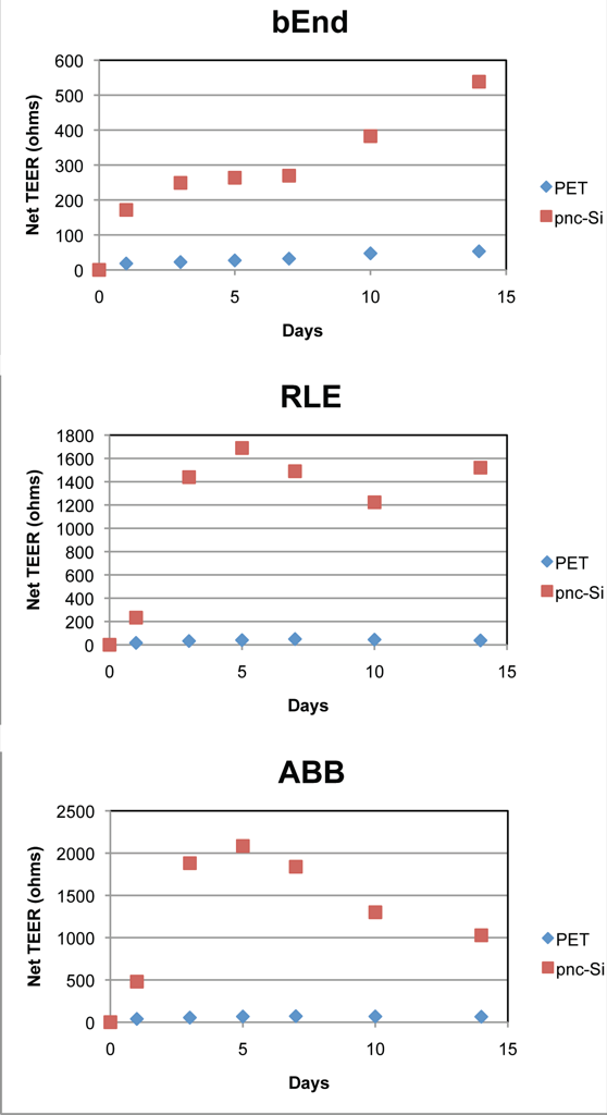
The following 3 graphs show the net increase in ohms (Day x TEER – Day 0 TEER). These graphs clearly demonstrate that the absolute increase in ohms is much greater for all cell types on pnc-Si than for PET. This is likely due to the smaller active area of pnc-Si than PET. There are a couple of reasons for this, probably. Edge effects (i.e., dead cells) manifest as lower TEER values for PET, which is not a problem for pnc-Si since pnc-Si slits are at the center of transwells. Also, TEER numbers for pnc-Si represent the barrier of only a couple hundred cells but TEER for PET includes the health of the entire monolayer. If there are dead cells scattered around the monolayer (almost always true), TEER would be compromised for PET but not necessarily for pnc-Si. This indicates that pnc-Si should be a better platform to detect subtle changes in TEER since the ohms values are so much higher than on PET.
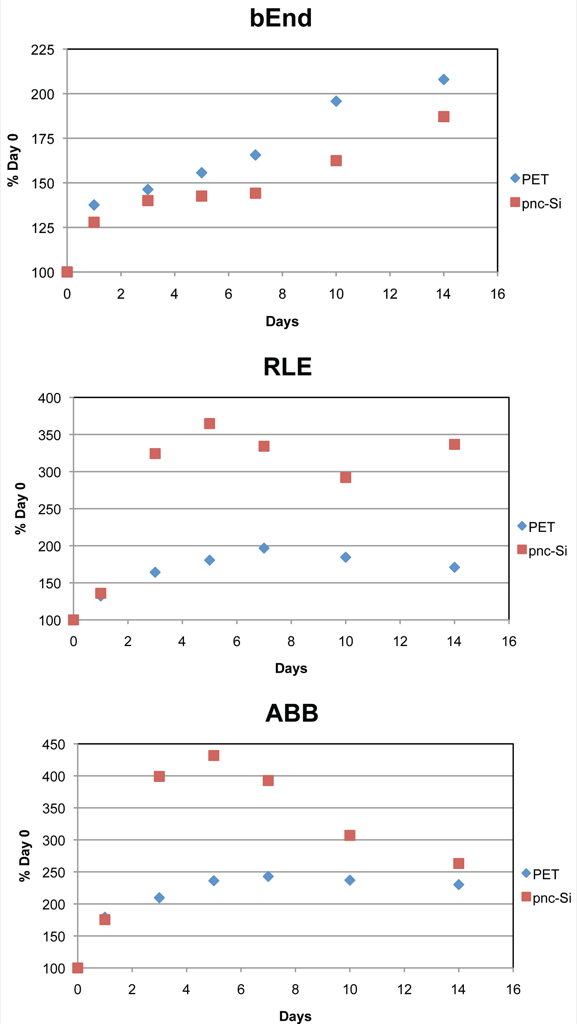
These graphs show basically the same data but normalized to be the % increase in TEER from day 0 values. Except for bEnd3 alone, there is a more rapid increase in TEER on pnc-Si compared to PET. This is also due to the smaller active area – it simply takes longer for cells to become confluent over the larger area of PET (including the edges).
These 2 graphs compare the cultures on each type of transwell (n=3, error bars are standard errors). Several things to note: The co-culture TEER was additive, so these cells types did not signal each other for tighter barrier function. The % increase in TEER on pnc-Si was higher than on PET (except for bEnd alone). There was a transient spike of extremely high TEER for RLE and co-cultures on pnc-Si. This is the same phenomenon that I observed with BBB co-cultures on pnc-Si. I think this is due to cell clumping by RLE-6TN (more on that later when I post Live/Dead images of these samples).
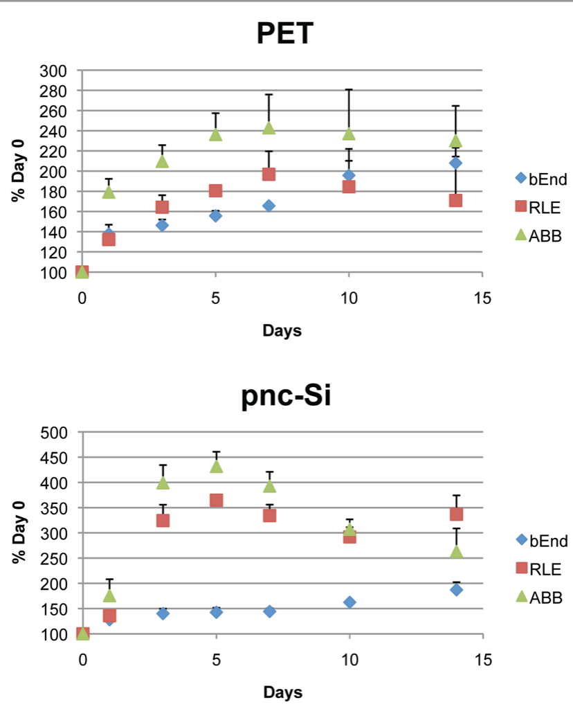
In conclusion, these results show that cells establish better barrier function more quickly on pnc-Si than PET. Also, co-cultures of these cell types do not result in synergistic increases in TEER. Last, the net increase (measured in ohms) in barrier function is ~100X higher on pnc-Si than PET.