Human Umbilical Vein Endothelial Cell Shear on Manufactured Hybrid (Dual-Scale) Membranes
Introduction
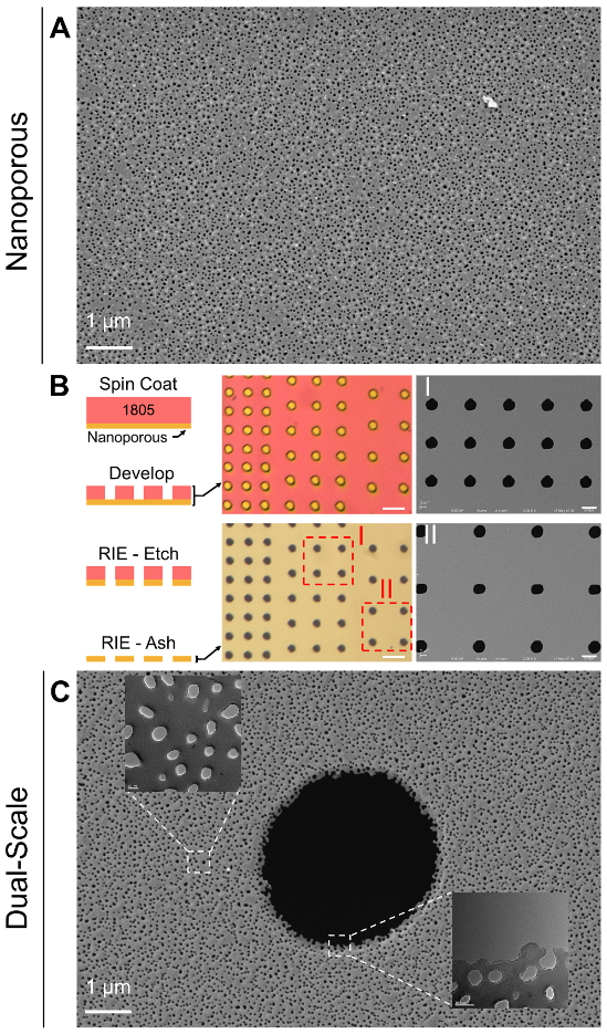
In our labs previously work, difficulties in growing endothelial cells (ECs) under shear flow lead to the exploration of novel membrane technologies that permit firm EC attachment, without compromising membrane permeability or leukocyte transmigration studies. We ultimately landed on the development of effectively ‘dual-scale’ membranes that contained 3 μm pores dispersed amongst an NPN (i.e. nanoporous) backdrop (Figure 1). Given the unique pore structure, the membranes retained high porosity values, regardless of micropore density. Using these experimental lithography techniques, we developed an array of varying micropore densities that permitted the elucidation of promising membrane candidates for shear flow and leukocyte migration studies. Recently, SiMPore took the concept and made it manufacturable on a wafer scale (Figure 2; 300-400 chips per run versus 10-20 previously). Thus, the experiments performed herein were to test biocompatibility (i.e. static culture) and cell adherence under flow as performed in the Dual-Scale publication.

Relevant Blog Posts
Dual Scale Membrane Fabrication
Dual Scale Flow and PMN Transmigration
Methods
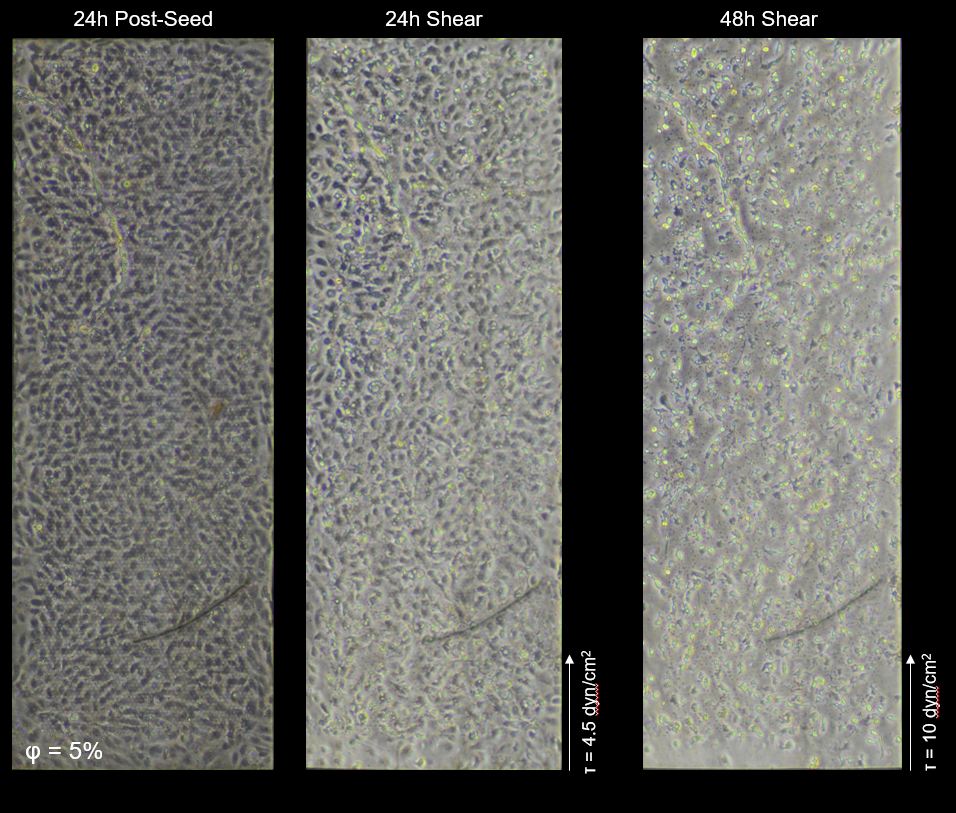
Human umbilical vein endothelial cells (HUVECs; Vec Technologies) were grown on fibronectin (Corning) coated (5 ug/cm^2; membranes coated for 1hr at RT, excess fibronectin washed away prior to cell addition) hybrid membranes. Following 24h static culture, devices were hooked to a flow circuit and sheared at 4.5 – 10 dyn/cm^2 for an additional 24-48h. Phase contrast microscopy images were collected at 0, 24, and 48h shear flow.
Membranes:
- 2.5% Porosity Hybrid (Dual-Scale), Wafer #1371
- 5% Porosity Hybrid (Dual-Scale), Wafer #1370
Results
In their first round of production, SiMPore manufactured dual-scale membranes with micropore porosities equal to 2.5 and 5%. Based on our prior work, the 5% micropore porosity dual-scale membranes were expected permit cell adherence under flow, but suffer from pore phase imaging. The 2.5% porosity membranes should also permit shear flow with the added benefit of clearer phase imaging. Shear flow experiments were repeated on SiMPore dual-scale membranes to test reproducibility of these results. On 2.5% porosity membranes, biocompatibility and clear phase imaging was observed over 24h HUVEC static cultures (Figure 3). Additionally, HUVECs remained adhered under shear flow (10 dyn/cm^2), and appear to begin polarizing to the flow direction.

Similarly, 5% dual-scale membranes showed biocompatibility, however, phase imaging is clearly affected by the abundance of micropores (Figure 4). HUVECs remained adhered under 4.5 dyn/cm^2 shear (original shear value used), but detached when shear levels were raised to 10 dyn/cm^2.
Conclusion
New SiMPore dual-scale membranes permitted shear priming of ECs under varying shear flow rates. This is not a completely unexpected result as these membranes are very similar in make to the originals (100 nm thickness, 3 um pores, 5 and 2.5% porosity). However, one important difference is the packing of the micropores: micropores were square packed originally, and hexagonally packed in the SiMPore variant. This is a subtle change, but not insignificant when relating to cell adherence. Nonetheless, the results are promising enough to move forward with the use of these membranes.
Nice work! Glad to see improvement of the circularity of the micropores with a proper lithographic tool. Do we have any TEMs of the material, or only STEM? Is there edge thinning (like we saw with the laser writer), and is there more of it with the thicker 100 nm material?
I agree! The pore patterning is much nicer with this fabrication process. We only have the STEMs for now, but definitely a good idea to collect TEMs at some point. Once we do that we can comment more on the edge thinning.