Carbonization of pnc-Si shrinks pores
After some experimentation with acetylene concentration and anneal temperature, I was able to find a carbonization recipe that appears to shrink our pores by 1/3 the original diameter. Below are images of an untreated sample (TEM 148: 15 nm pnc-Si 1000 RTP) and a carbonized sample (see recipe at end of post).
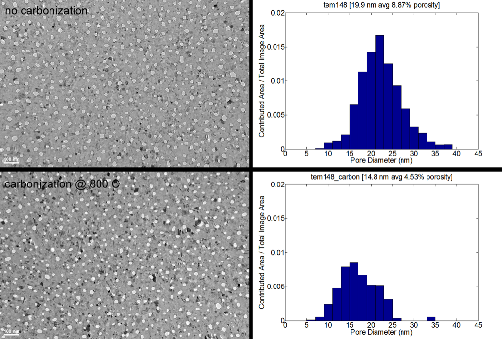
A closer look at the carbonized sample shows a “ring” of material around the pore. Previous studies with carbonization on porous silicon suggests that the reduction in pore size is caused by the formation of a layer of SiC. I did not notice any wrinkling in the film after treatment.
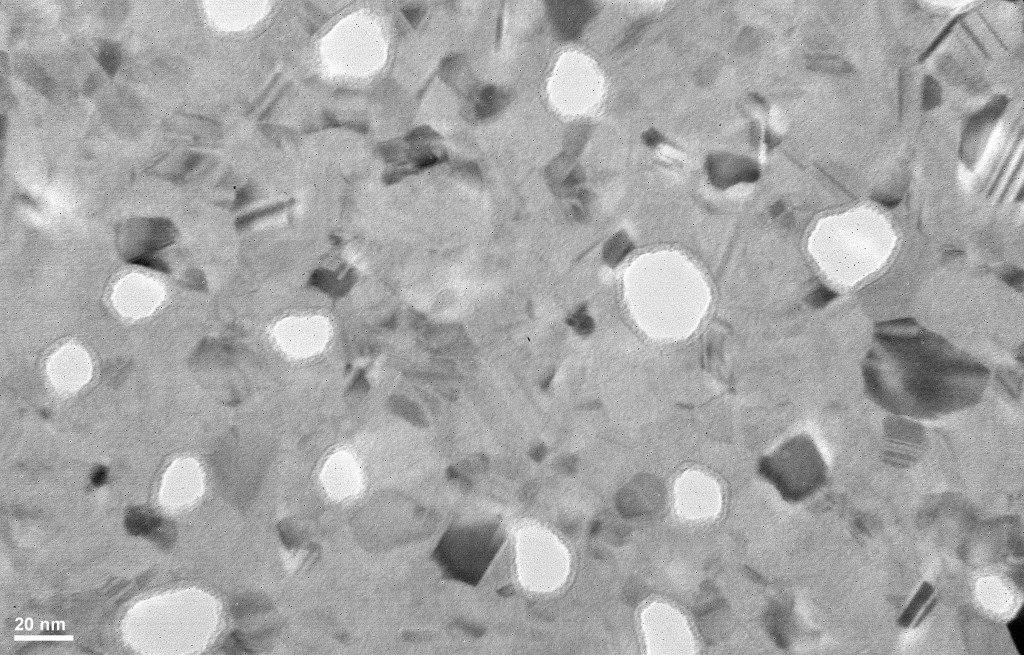
Here are two high resolution images taken on Brian’s scope:
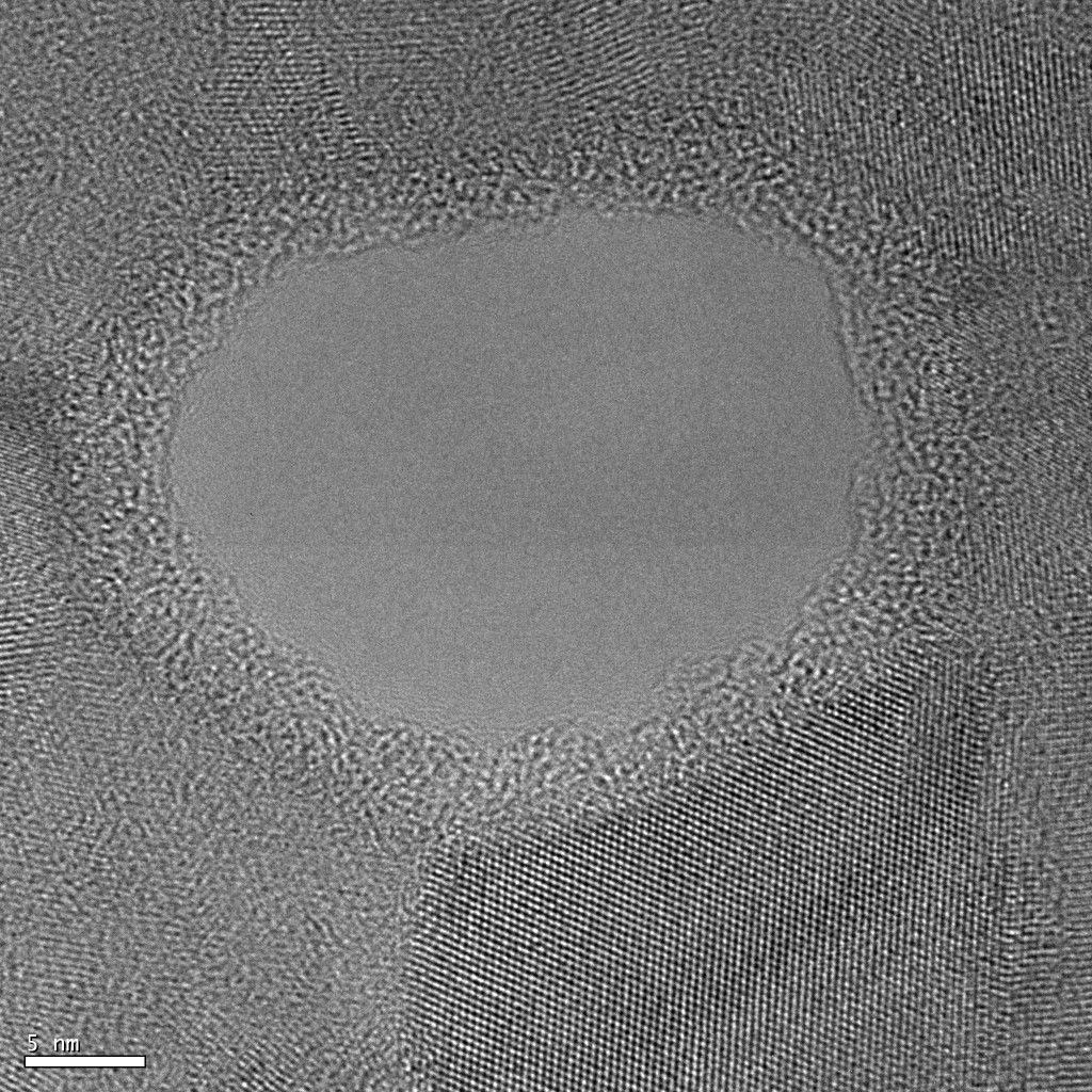
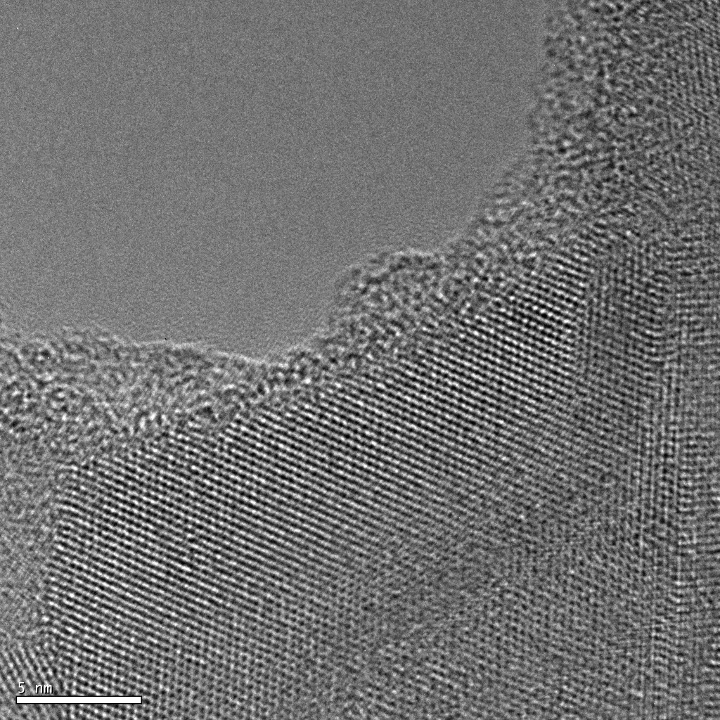
There is a clear distinction between the nanocrystal and the amorphous carbon border.
I was curious to see if we could pick up a carbon signature using scanning EDX spectroscopy. Below is a scan over a single pore.
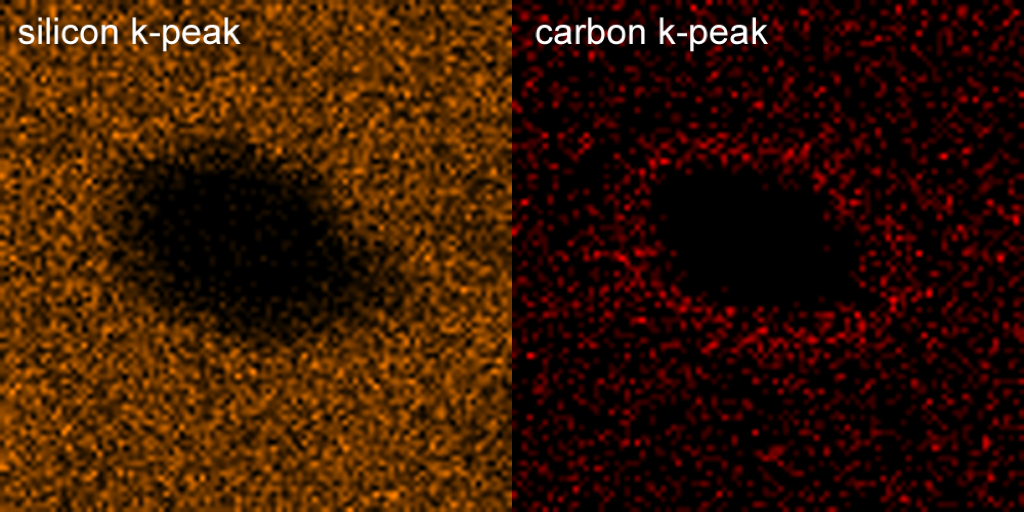
As expected, EDX picked up a strong silicon signal everywhere except for the pore. More interestingly is the carbon signal which is most concentrated around the pore. This means that we are indeed closing the pores with Si:C.
The recipe I used was pretty aggresive: 20% acetylene at 800 C with no dwell time between the acetylene flush and thermal treatment. Literature suggests that the thickness of the SiC layer can be tuned by delaying the thermal treatment after the acetylene flush.
Here’s the protocol I used:
- Dip membrane in 50:1 HF for 60 s, rinse and dry – important because we need Si-H terminations
- Immediately put in RTP on top of susceptor (uncovered)
- Flush RTP chamber with 10 LPM Ar for 5 min at 20 C
- Flush RTP chamber with 8 LPM Ar and 2 LPM C2H2 for 5 min at 20 C
- Heat chamber to 800 C with 10 LPM Ar for 5 min
- Cool chamber down for 10 min with 10 LPM Ar
Now that we’ve confirmed a change in morphology, further characterization needs to be performed using contact angle measurements and discoloration tests.
this result is very nice, Dave. This gives us a practical handle for tayloring the pore size. The TEM images clearly show that you have an inner ring of a different material- could we do HREELS or something else to measure the compisition of this material? Also, could permeability studies be done of the sample before and after (let’s do air to avoid possible chemistry issues)- this would be a very strong result to show….
I agree, this is very exciting. I tried to pick out the carbon using EDX but it’s too light. Do we know someone who has access to EELS?
Just found this: http://people.ccmr.cornell.edu/~davidm/Muller_facilities.html
I think we’ve collaborated with him before, right?
yes, we have collaborated with him. I think through SiMPore, so maybe Tom or Chris could do the initial “asking”
This is really cool and could be the basis for an interesting short paper. It is the first demonstration of Rapid Thermal Carbonization, correct?
In the 2 compared images, the one without carbonization appears to be at a different focus point than the one with carbonization. This may have consequences for the pore processing and interpretation, so for publication, we should try to keep the focus a little closer.
In the recipe, is there a specific reason for turning off the acetylene for the high temp step? This would then allow only that acetylene left in the chamber, to react with the surface. I think it would be better to keep the acetylene on at high temp, so the concentration is stable. Was the susceptor used?
Did you put a drop of water on the sample to determine if it is hydrophobic? I’m curious.
This is a good first step. I know Muller, but it takes forever to get anything done. We should try EDX in Brian’s TEM first, and also get a little more experience with the process.
If you want discoloration studies done, bring me some samples (if you can spare any) to the meeting on Tuesday.
Very very nice. If we tighten-up the carbonization procedure so that only the temperature or the acetylene flow*time are variable, I think this is ready for publication – maybe Nano Letters or ACS Nano? This is plenty for an initial show and tell letter, then follow-up with a more detailed paper that includes dissolution studies, contact angle, cutoff tuning for protein/DNA, etc.