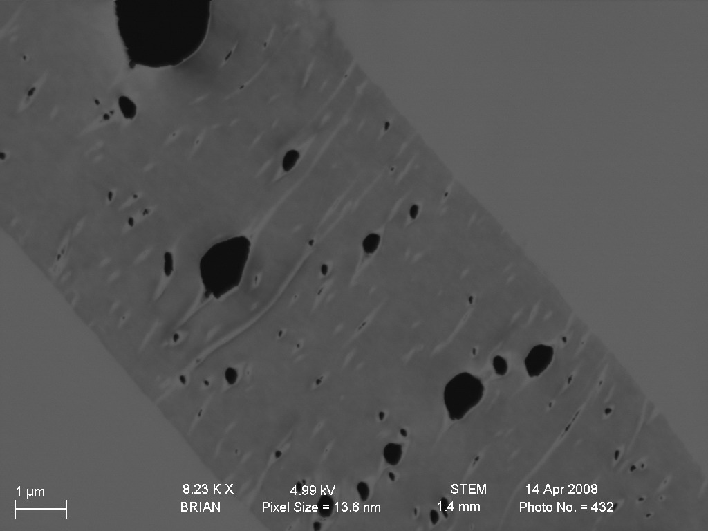
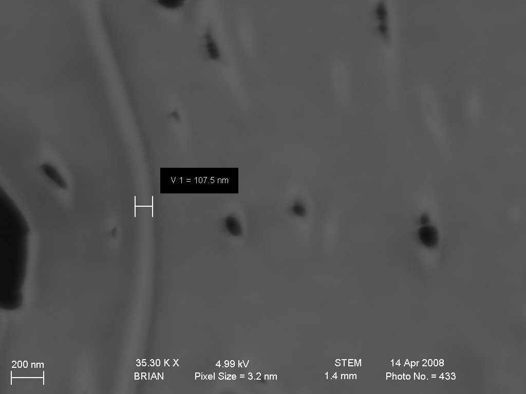
SEM images of vertically cut track-etched membranes
TE membranes were first embedded in plastic material and then these samples were cut with microtome and imaged. The images below are of the 30nm and 80nm pore diameter membranes.
30nm ones came out better. It looks like the diameter isn’t really 30nm, look at the second picture. The dark spots are either tears or voids that were etched during processing.


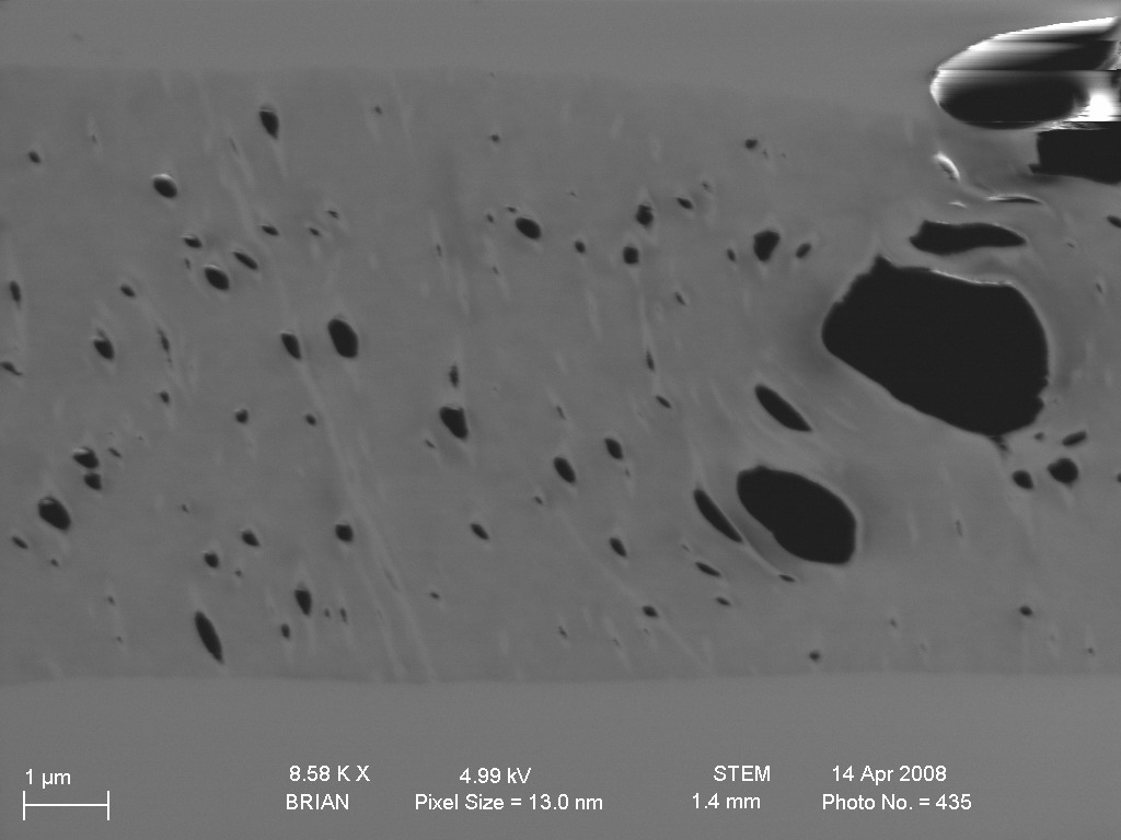

Also two 80nm diameter membranes images. They are not very clear also I expected it to be much better then the previous ones.


These images show that the pores are not straight and probably are of different diameters through the microns of the polymer. It confirms difference between theoretical and experimental air permeability numbers for TE membranes.
I think you should try taking the microtome and taking slices at a 45 deg angle, instead of a true 90 deg cross-section. This will allow you to see high-contrast holes, instead of the gray streaks viewed here. Some of the 30nm pics show some holes that are close to the correct size, but a 45 deg image should give you many more pores to measure.
These are very nice preliminary images – what does the surface look like? Can you see pores?