BSA detection on NC
Creating an array to hold a pnc-Si wafer to proving to be difficult. The PDMS spreads too rapidly though the NC to form a consistent array for the pnc-Si membrane (and eventually wafer) to rest on. I’m still running different trials of the PDMS on NC to see if I can get the array to work with a different ratio of Elastomer base: curing agent, and also using different methods of application.
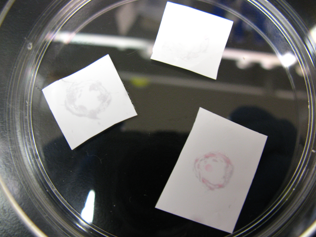
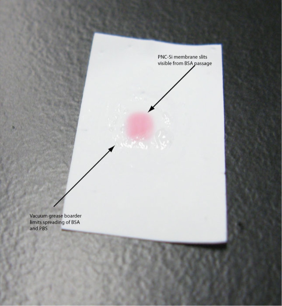
To show that NC and Ponceau S can be used as a detection method for BSA, I set up a system with pnc-Si on a NC, suspended by vacuum grease + Si beads. The membranes used for these trials were from W677 (the one that doesn’t fit in the Sep-Cons). The backside of the membrane was wet with 15uL of1X PBS. I also pipetted 15uL of PBS onto the NC to pre-wet the NC membrane. The wells of the pnc-Si membrane were filled with 5uL of 1mg/mL BSA. Before putting the pnc-Si on the NC, I outlined a circle, slightly larger than the pnc-Si on the NC paper using vacuum grease to prevent spreading. This seems to work pretty well, however, some spreading still occurs. The picture below (on the left) is the first trial of BSA on the membrane system, after it was stained with Ponceau S. The experiment ran for almost 24 hours (+/- 2hours). The pnc-Si membrane was carefully removed, attention was paid as to not spill the remaining BSA off the top of the membrane. The NC was rinsed in 1X PBS for 1 minute to remove any residual BSA from the NC, then the membrane was stained with Ponceau S. The experiment was repeated on Tuesday, with the result on the right. However, this time the stain was much lighter. It is possible that the Ponceau S was rinsed more thoroughly the second time, since Harold said that shade of the dye with respect to the concentration of the protein is not quantifiable.
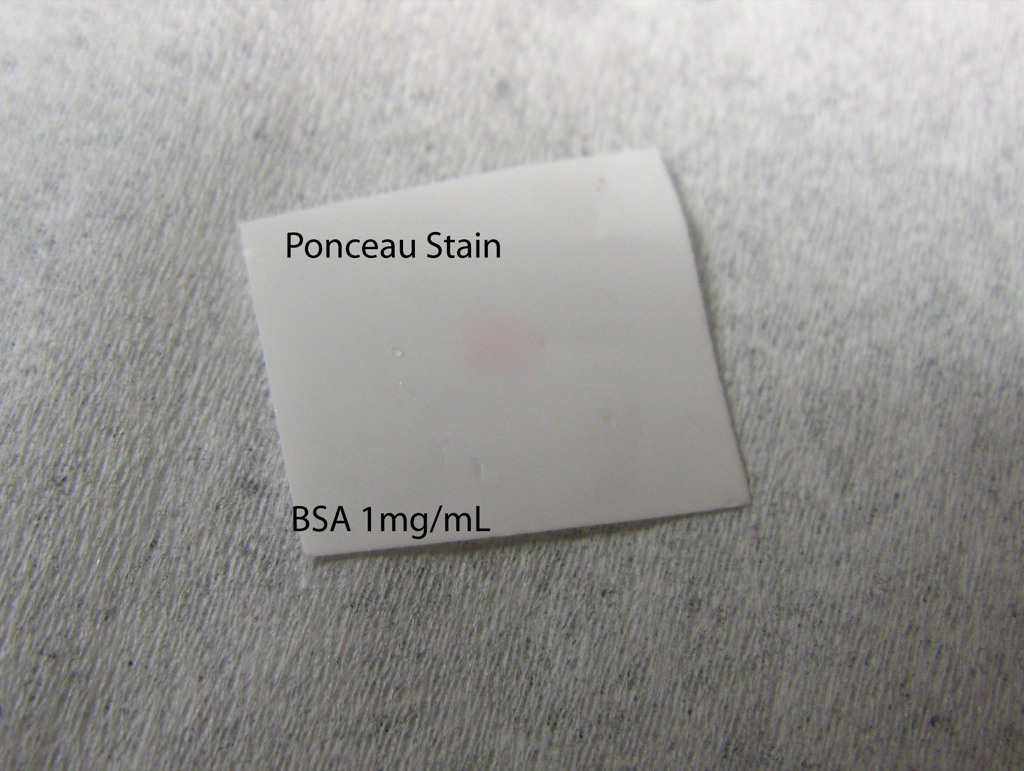
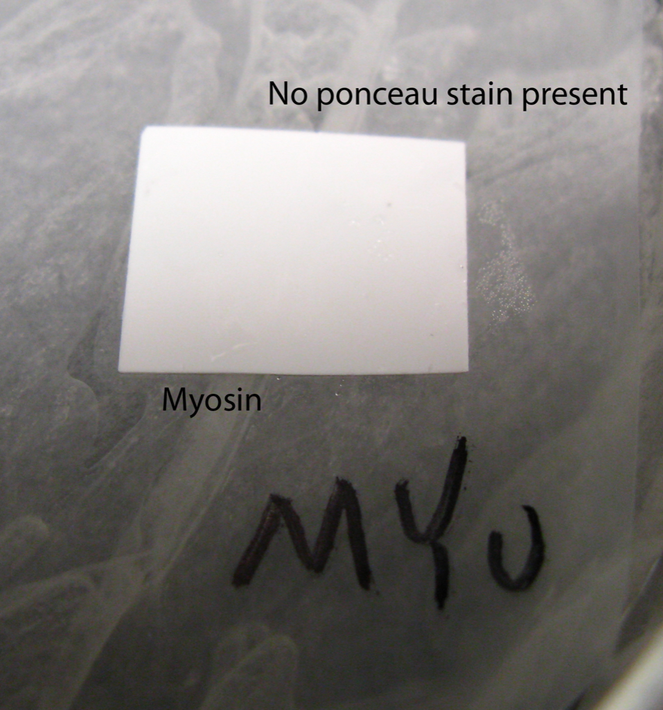
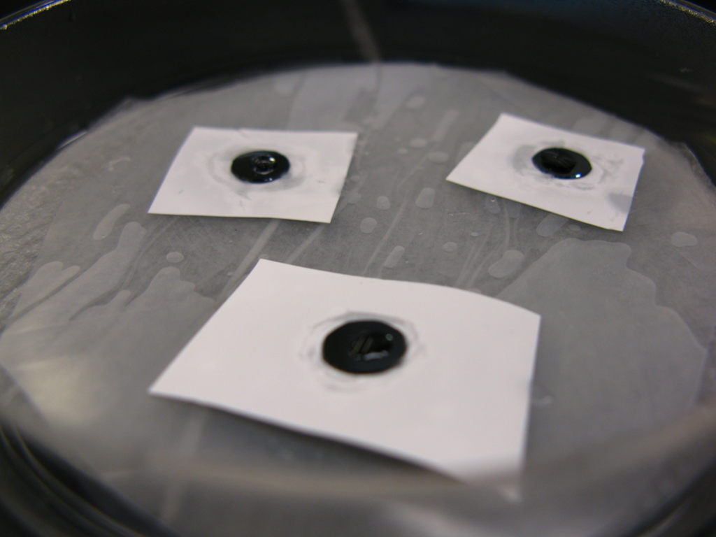
Along with attempting to diffuse BSA through pnc-Si onto NC, I ran the experiment simultaneously with myosin, since we know that myosin does not go through out membranes. This is illustrated below in the image to the left. On the right is an image of BSA, Myosin and Myosin + BSA on the NC in a humidity chamber. The results of this experiment were quite odd. The BSA did not show any diffusion, but the BSA + Myosin (NC membrane in the lower right of the image) showed a small pink dot on the NC.