Discoloration – SepCon format
I performed a discoloration experiment with w639 in EGM + 10% FBS. I put 2 blank chips in the Sepcons membrane side down. I then placed one Sepcon in a well with EGM alone. The other Sepcon was placed in a well with EGM but also with another chip (labelled as Sepcon with chip, chip with Sepcon). The control was a chip in EGM alone. This was done in the incubator.
Here’s a quick pic of the experimental setup:
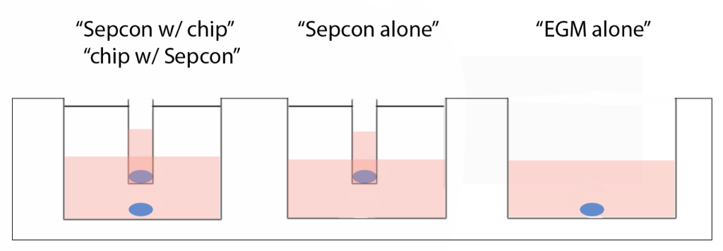
By using blank chips, I could isolate the “well side” from the membrane side of the chip. If the “well side” catalyzes discoloration, then discoloration in Sepcons should be slower. Alternatively, something about the Sepcon format could be preventing discoloration (something leaching from the O-ring or plastic housing?). By placing a chip in the well below the Sepcon, I thought that the “well side” of this chip could catalyze the discoloration of both itself and the chip in the SepCon format.
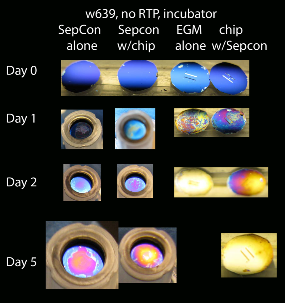
After 2 days, the chip in EGM alone has totally discolored, but neither of the chips in the Sepcon format are really discolored (I think the slight purple color comes from problems I had in rinsing and effectively drying the chips in this format). The chip in the well with the Sepcon is slightly less discolored than the chip by itself. Could this be because the backside of that chip was catalyzing the discoloration of 2 chips instead of 1? After 5 days, the chip in the Sepcon by itself has barely discolored. However, the chip in the Sepcon that had also been exposed to a chip in the well was discolored.
This result suggests that there is nothing about the Sepcon format (i.e., plasticizers leaching from the components) that prevents discoloration. I think this is pretty convincing evidence that the “well side” plays a role in discoloration.
Looks good. It will be interesting to see how the bead assay comes out to be in the sepcon format. That should tell us whether the well side plays a role in the actual membrane degradation or the membrane is attacked directly.
I used cloning rings in most of the experiments (for cell growth, adhesion studies) and there also the membrane side was separated from the well side like in the sepcon format. But discoloration did take place as it does usually. So how can this be explained in such a context? May be the separation using cloning rings and silicone was not as strong as due to sepcon.
Good point Anant.
I suppose it’s possible that the seal with cloning rings isn’t as tight as Sepcons, but I find that a bit hard to believe. With cloning rings, the pnc-Si is still flat. This points to something unique about flat vs. suspended in a Sepcon, or something about the Sepcon itself.
Maybe the SepCon exudes some kind of plastic molecule that protects the pnc-Si. What are the cloning rings made from?
Cloning rings are made of polystyrene.