TEM images of pnc-Si incubated in DMEM(-FBS)
There has been uncertainty with process of dissolution of pnc-Si in cell growth media. Whether the media enters the pores and ‘eats away’ the material leading to increase in pore sizes and eventually complete degradation was the question. Thinning of the material would have taken place too. To have a clear picture about this, I wanted to look at the change in TEM images of the material and observe if there is any change in pore features over incubation time in cell growth media.
For this, I took samples from W673 (high porosity; used for permeability experiments) and incubated them in DMEM(-FBS) in oven @ 37C. These samples were not treated with anything. Samples were then taken out from media at different time points of 90min, 150min, 6h and 24h (t=0 sample was not dipped at all) and rinsed with dH2O and IPA followed by N2 drying. t=0, 90 and 150 min samples had intact membranes (with no sign of discoloration) and thus were imaged using TEM by Dave. Whereas, the 6h sample turned golden and its membrane had degraded. Similarly, the 24h sample turned silver with no free standing membrane. All the samples could be imaged using AFM to see the change in porosity (as Barrett can do AFM on the supported as well as free standing membrane).
Following are the captured images:
1) t=0 (100000X)
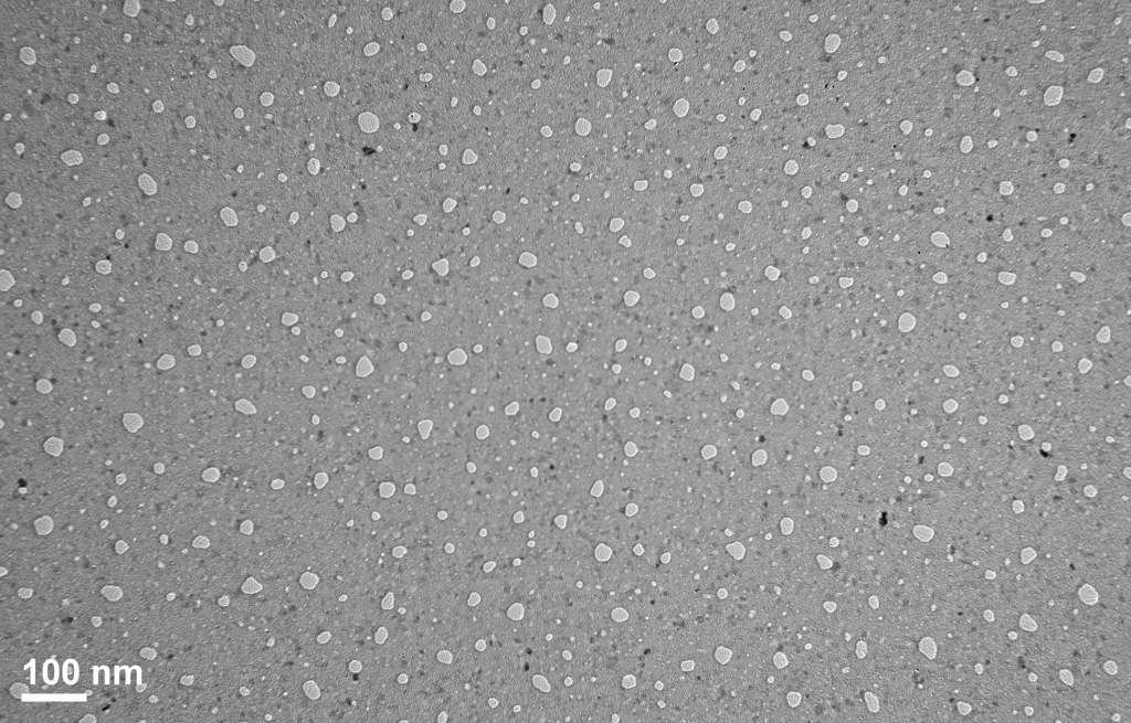
2) t=90 min (100000X)
There is not much visible difference between this and the above sample.
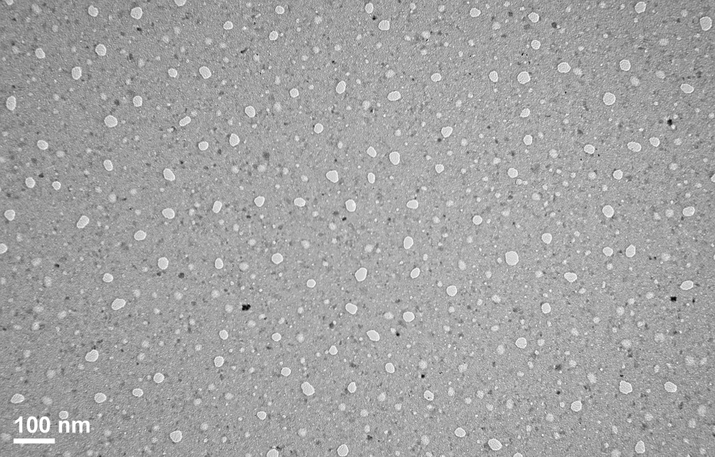
3) t=150 min (100000X)
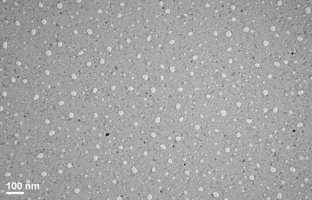
Here, it can be seen that the pores are comparatively bigger than the above images. Also, the pore boundaries appear to be irregular whereas the t=0 sample has pores with smooth boundary. Also with the intensity of light transmitted, the material appears to be thinner here. The black spots are just some stuff from the media.
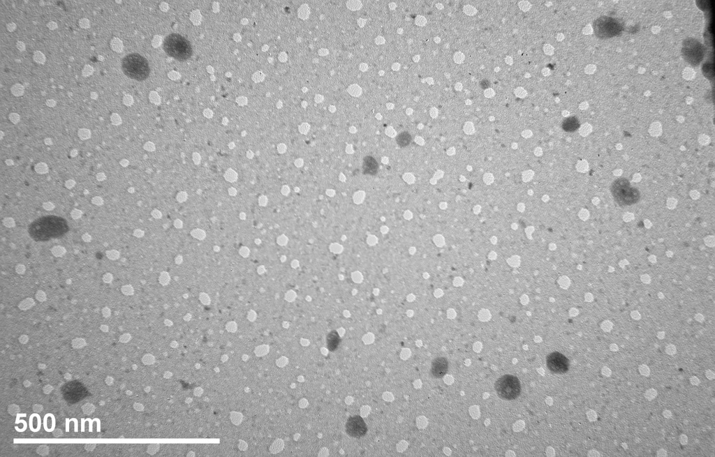
The following zoomed in image (200000X) gives a better look at the pores and it does seem that the media is eating away the pore boundaries.
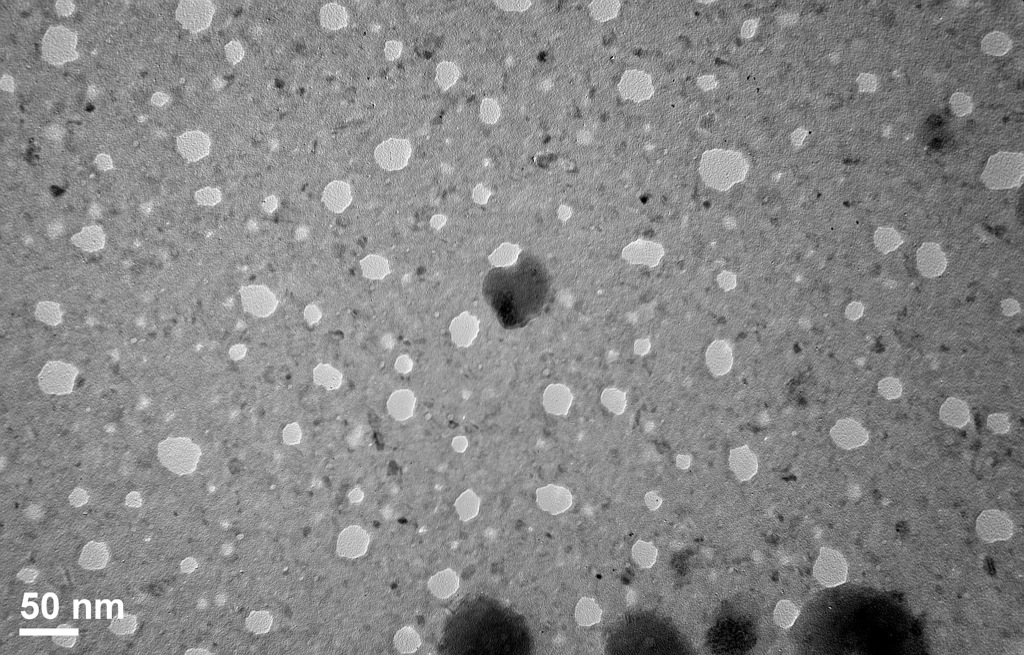
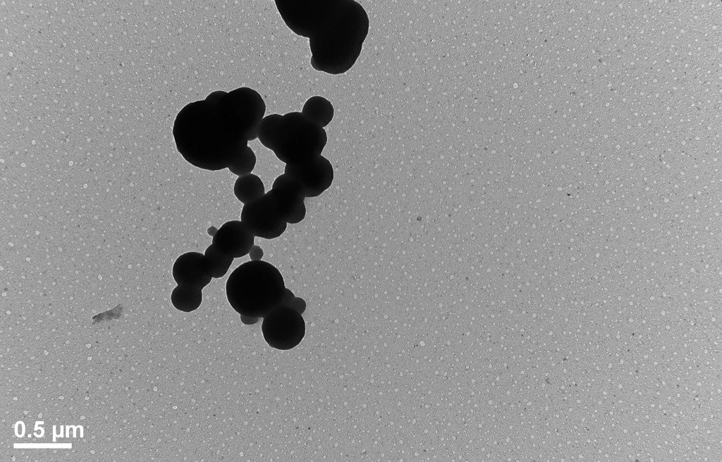
Pore Processing of these images can show the increase in cut off (highest pore size) with increasing incubation time. I will do that next. Also AFM images can confirm this. What you see below is some type of debris from the cell culture media; it could be yeast/molds. (30000X)
Nice. For me this is first direct evidence that the material is really dissolving rather than delaminating or cracking and flaking.