Day2:Cell Adhesion (Trial 3)
On Day 2 of cell growth from trial 3 of adhesion study, cells looked much more spread out but less in density. Most of the cells were observed to have concentrated near the edge of the pnc-Si chip near the Sepcon as seen in the figure below (first image in the bottom row). This could be due to the influx of media through the broken membrane that pushes the cells away. Or, this could have happened during the staining procedure.
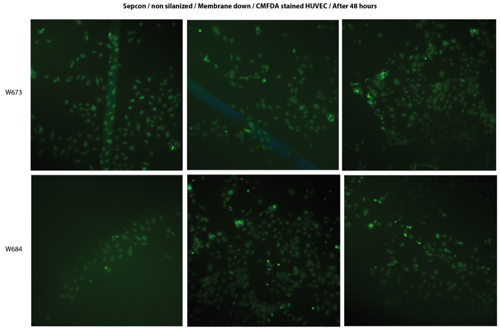
The whole purpose of this experiment is to observe the cells form a confluent monolayer which could give us an idea about the plating density of the cells to get confluency within the stability period of pncSi in 3D format.
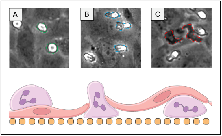
I’ve found that cell density is lower around broken slits in my fluorescent images also. I think it means that the slit is breaking during the staining protocol, which allows fluid to flow through the slit and shear off cells near the slit. Are these the same samples from day 1?
Yes, these are the same samples.
Also, that was day 0 (4-5 hours after plating).