Monolayers, not clumps on pnc-Si membrane
In my last couple of posts with respect to endothelial cell culture on pnc-Si transwells, I presented images of cell clumps over the active pnc-Si membrane area of transwells (here and here). There were dead cells associated with these clumps and this is a concern for barrier function experiments. This post shows a counter-example of cell clumps on pnc-Si.
These are P10 cells, seeded at 50000 cells/cm2 and grown for 14 days. I then stained the cells with Live/Dead, so live cells are green.
Phase contrast:
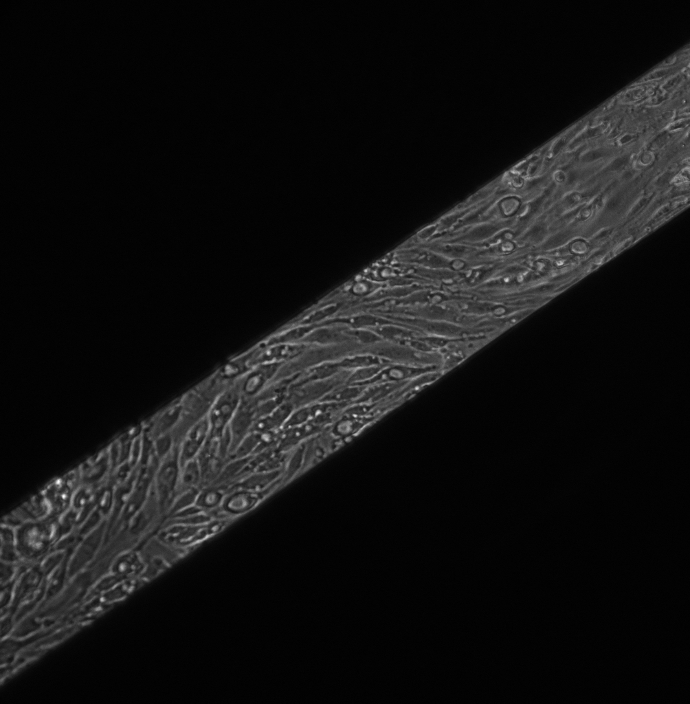
I haven’t enhanced this image at all and the contrast is excellent. I presented this image at BMES.
Fluorescent Live:
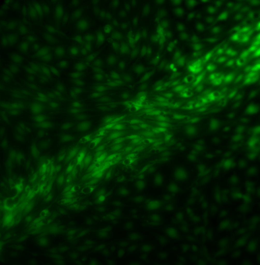
With the fluorescence, it is clear that there is a “clump-less” monolayer growing across the pnc-Si membrane. Interestingly, this sample is a 50nm thick, nearly pore-less membrane. I’m not sure if the thicker membrane or the low porosity could prevent formation of cell clumps.
If you look closely at this image, you can clearly see cell nuclei or other large organelles (black circles) in the cell only on the free-standing pnc-Si membrane. No nuclei are visible on the supported membrane. I’ve see this on other pnc-Si membranes, as well. Additionally, I’ve noticed this on PET transwells (see below – these are P8 cells grown for 14 days), although not all samples show this phenomenon on PET.
I’m looking at references to see what this staining artifact means. Maybe as the cells get older, more of their cytoplasm is taken up by large nuclei organelles? Maybe the cells flatten over time so that there is little cytoplasm to fluorescence on either side of these organelles/nuclei?
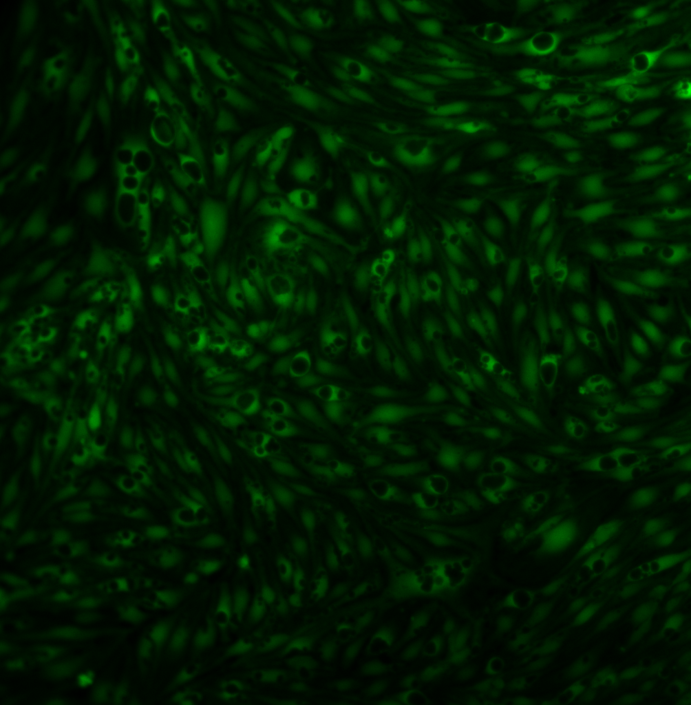
Notice the skull-looking cell in the upper left corner.

Happy early Halloween.
The black spots are vacuoles, not nuclei. Endothelial cells are famous for making these and they are important for transendothelial permeability. It is pretty interesting that these occur only on the membrane area although I don’t know what it means. We might want to show this figure to Ingrid Sarelius at Thursday’s PPG meeting.
The phase contrast image is just terrific.