HUVECs Contracting on Nonporous SiO2 120nm Membranes
While performing an experiment to study HUVEC growth rates on various substrates, I observed an interesting phenomena on the oxide membranes. I was using 120nm, nonporous, 600C annealed SiO2 membranes in this trial. The cells were observed at least once per day over a course of 173 hours from seeding. The growth rates and quantitative data are in this post.
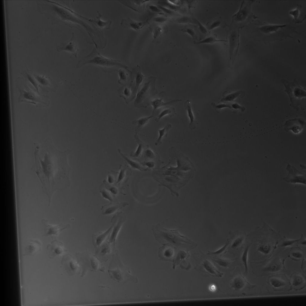
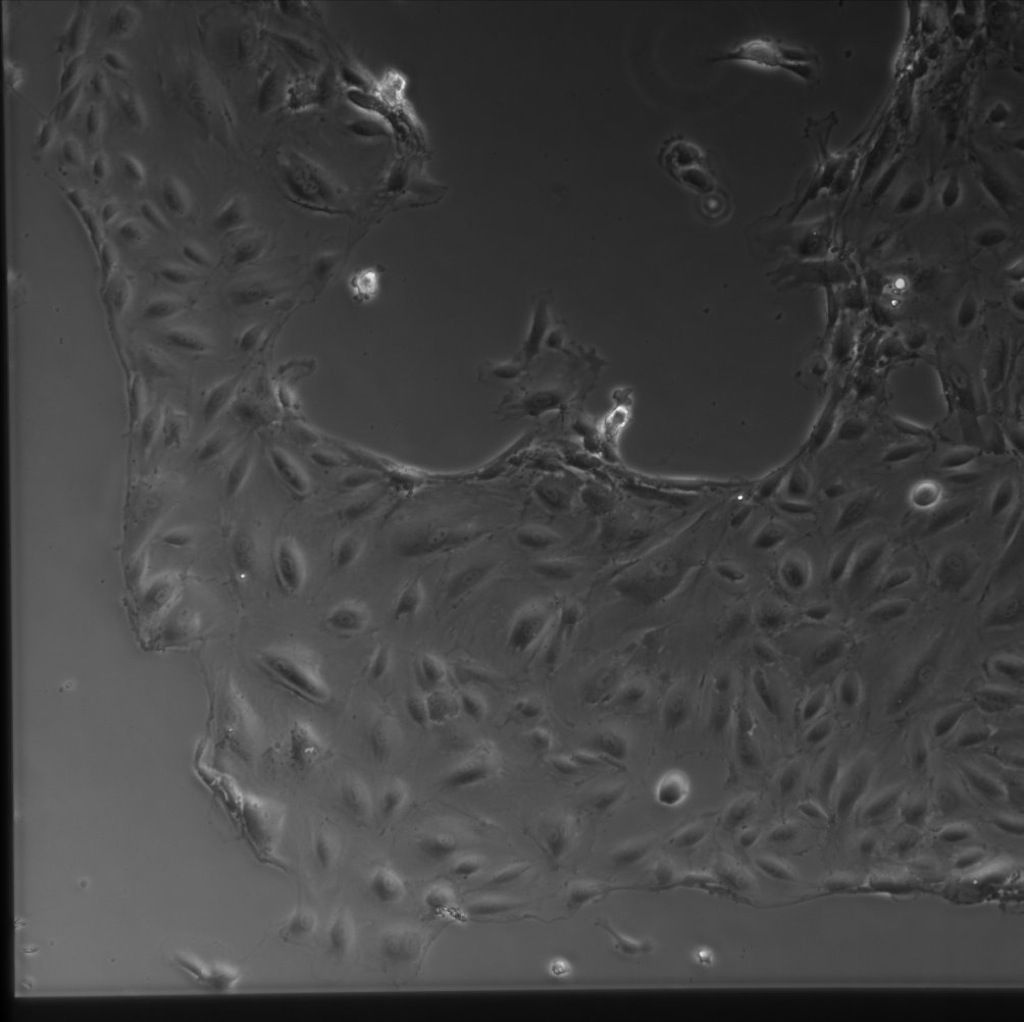
It appears that under some sort of stimuli on an oxide membrane during the experiment, the HUVECs stopped approaching confluence and, while remaining viable, began constricting into more narrow bands and ‘linings’.
The following images give an idea of the relative growth area available on each substrate:
TC Plastic (and glass) were constricted to a silicone gasket of ~14mm²:

The SiO2 membranes (and SiN) have ~4mm² of surface area to support cell growth:

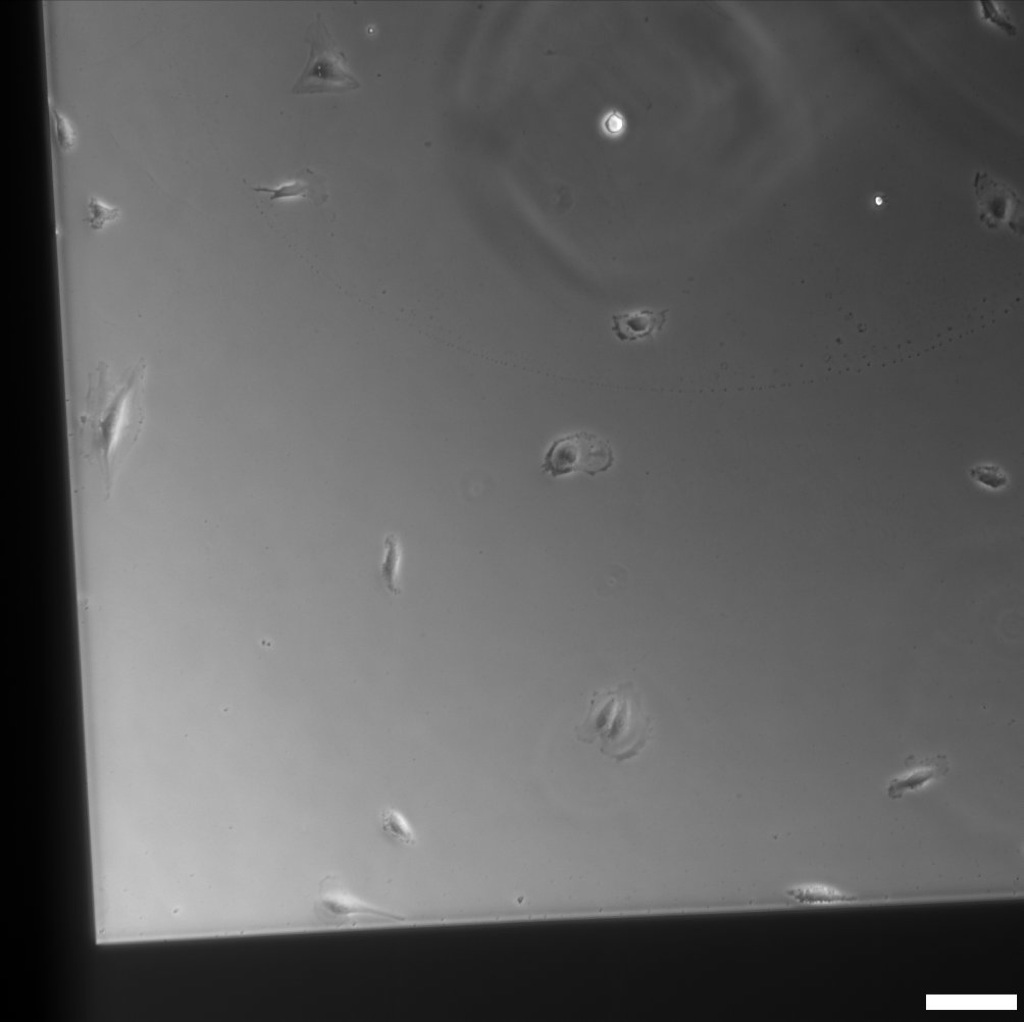
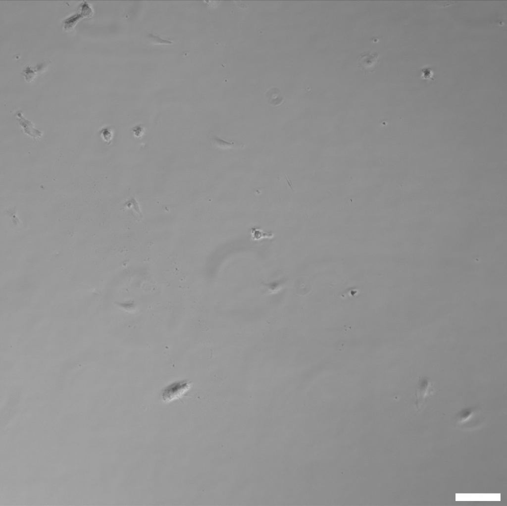
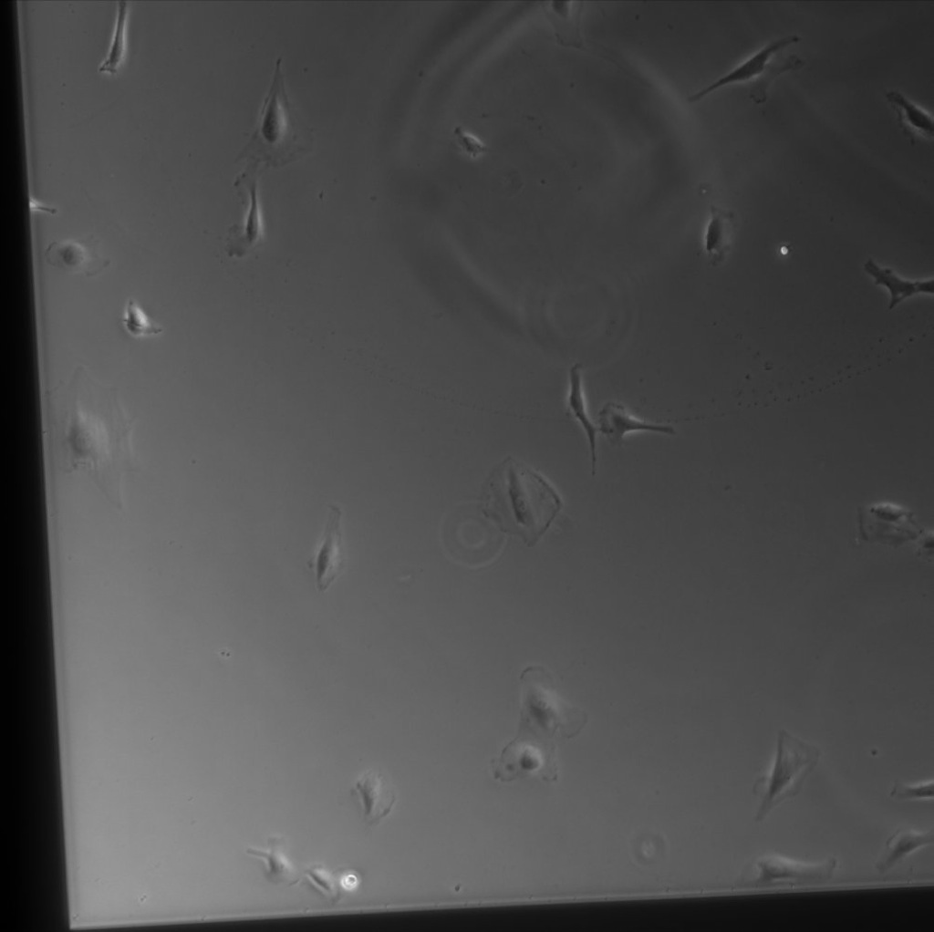
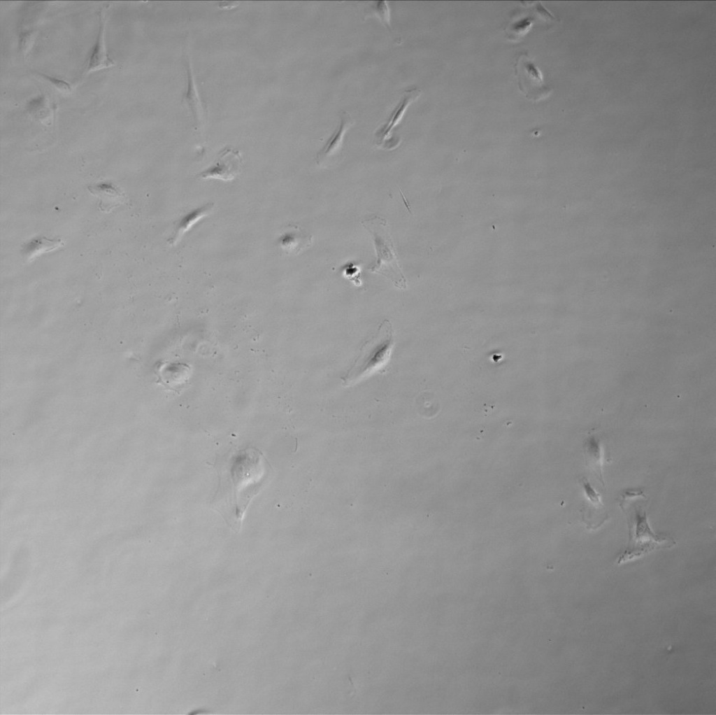
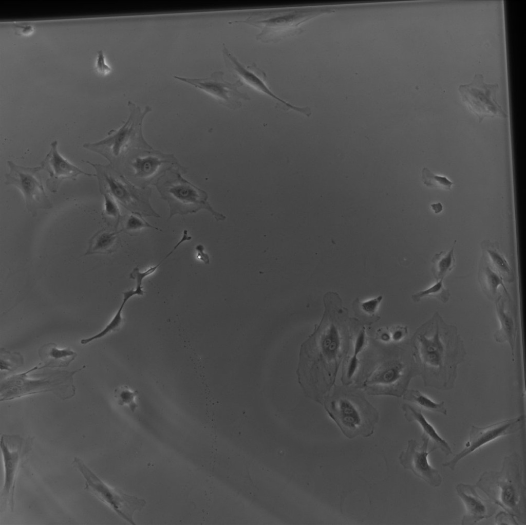
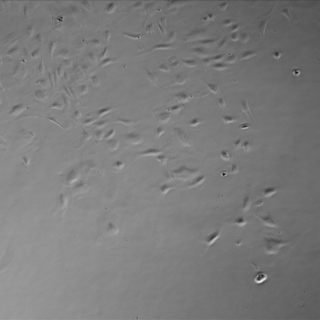
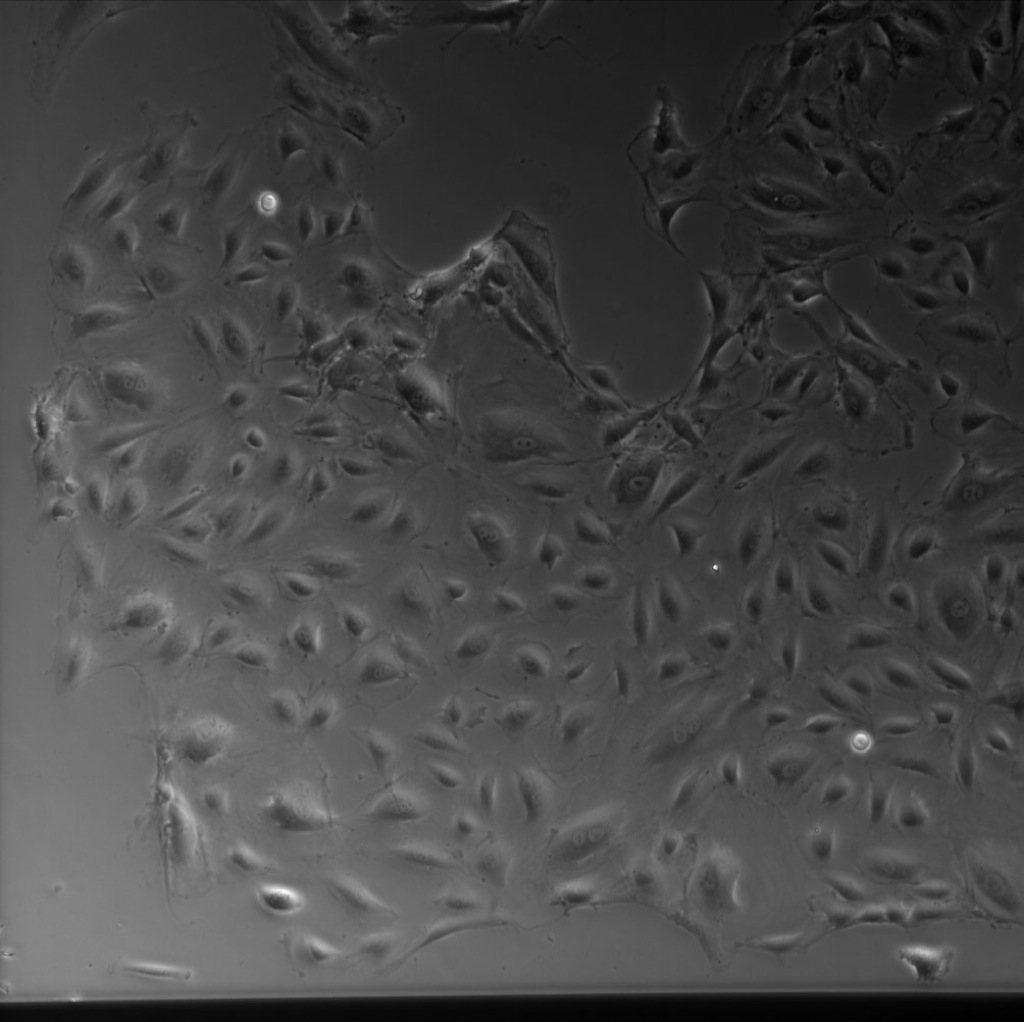
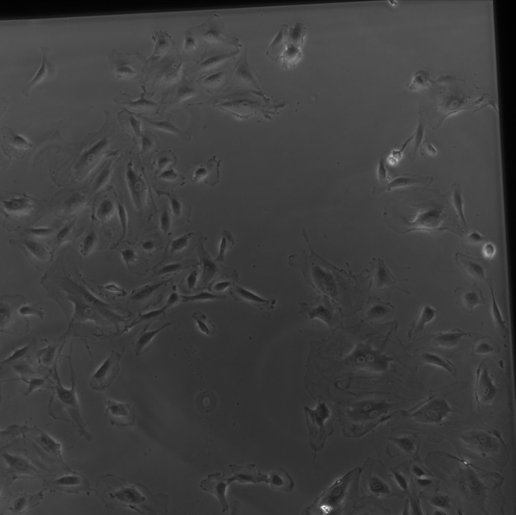
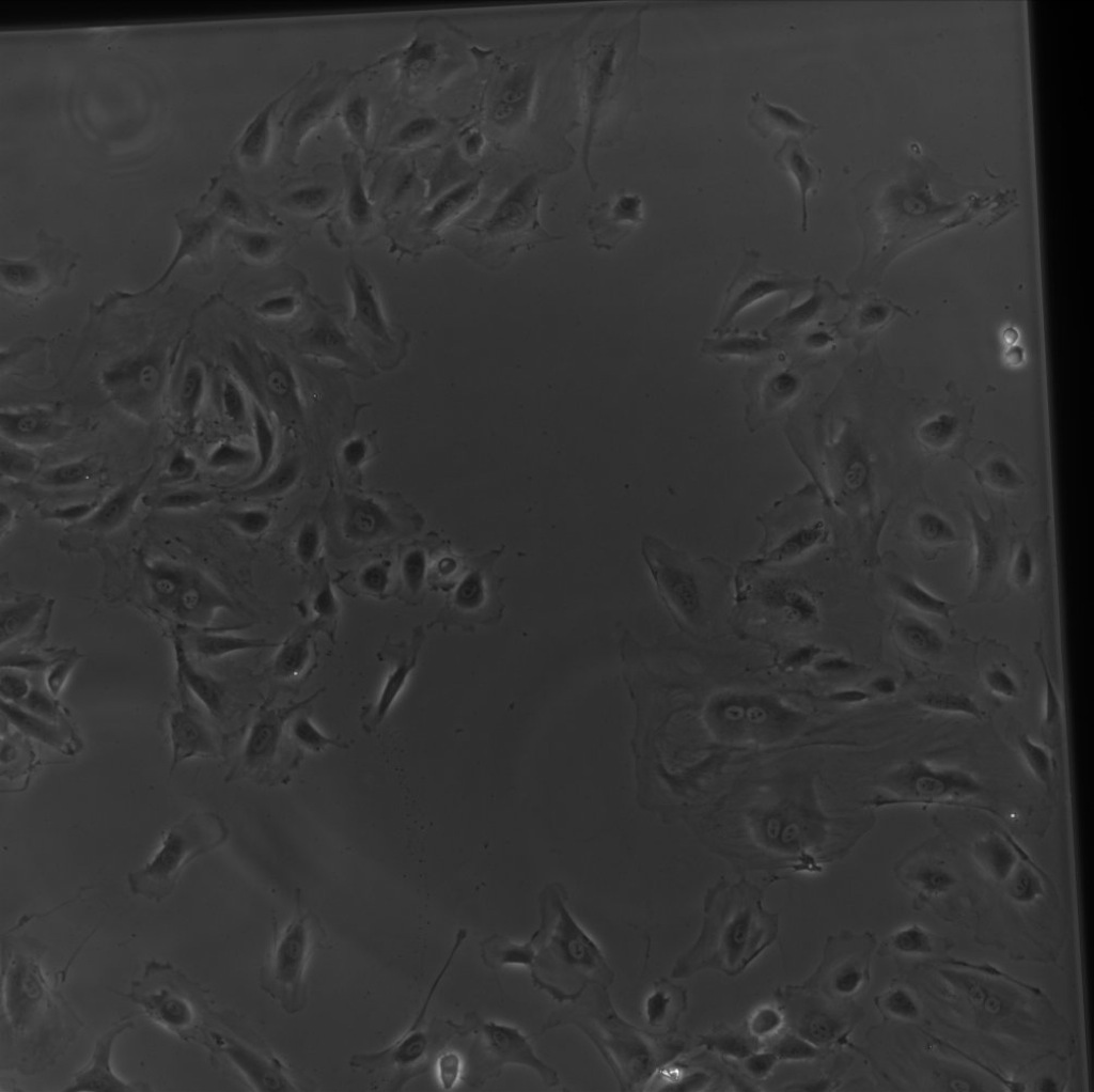
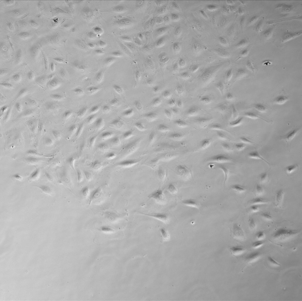
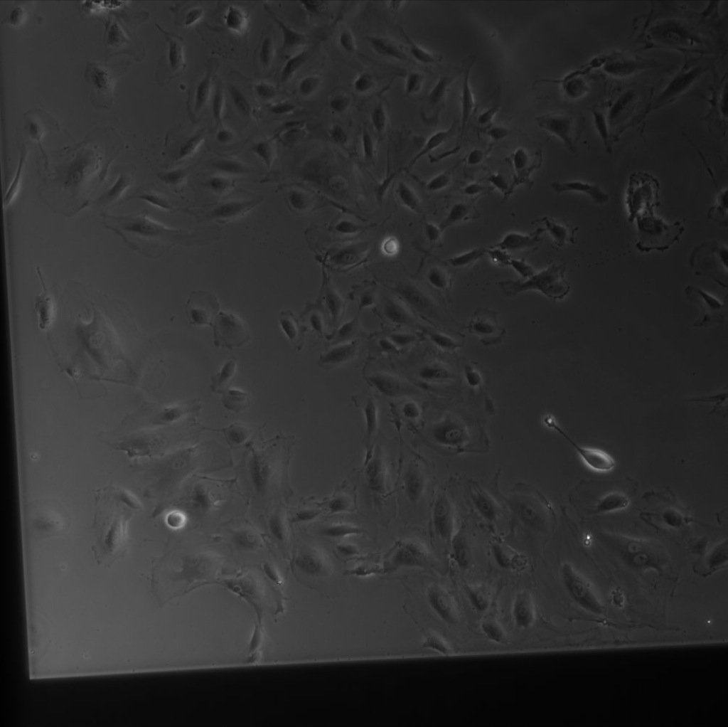
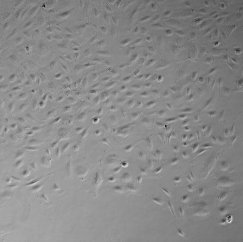
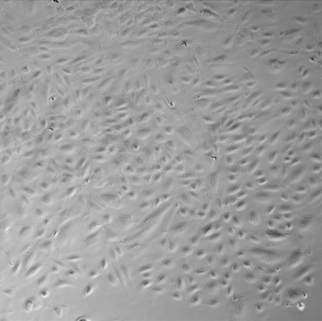
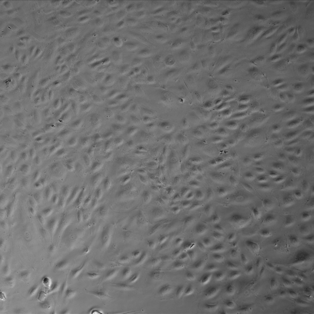
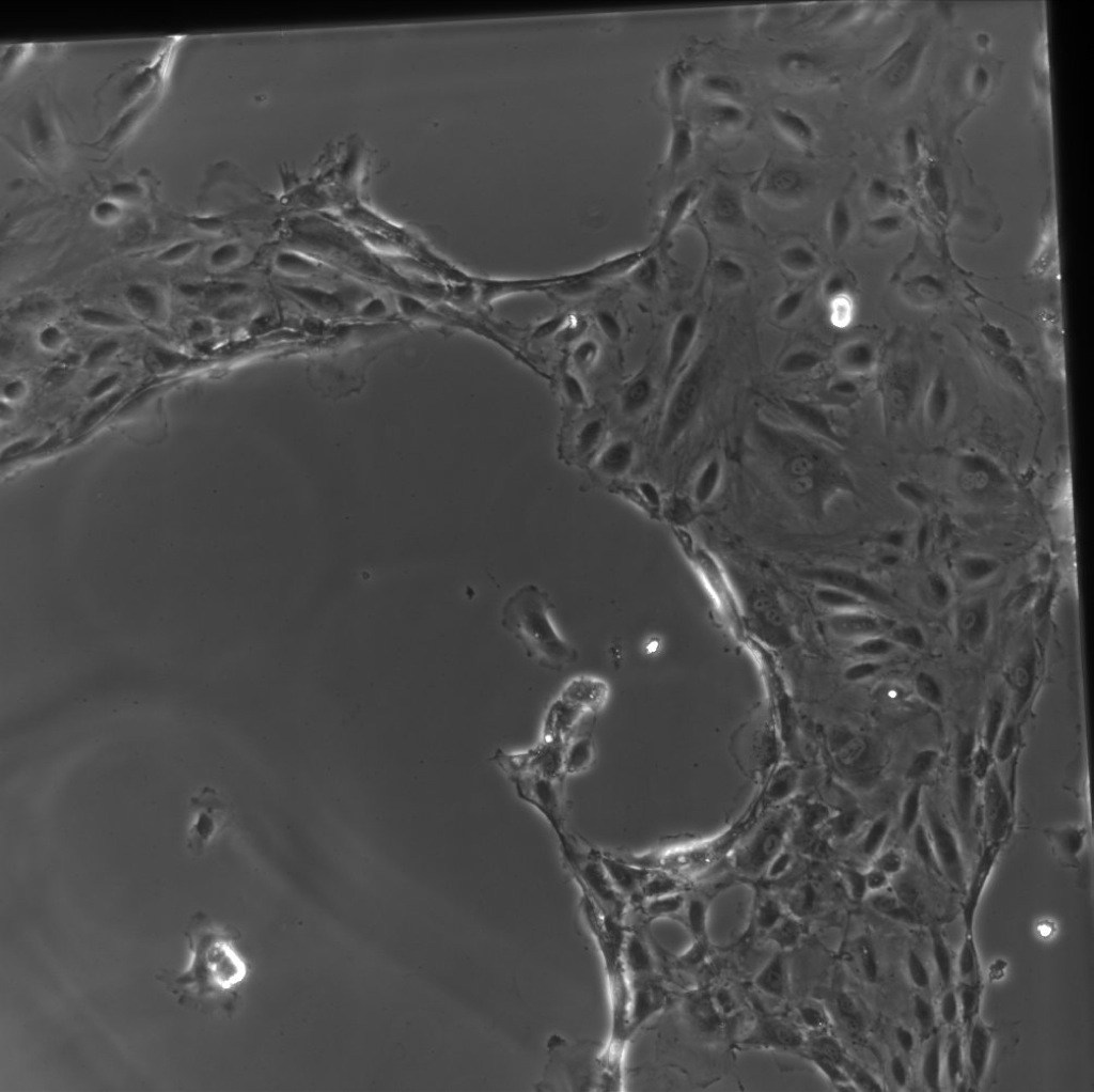
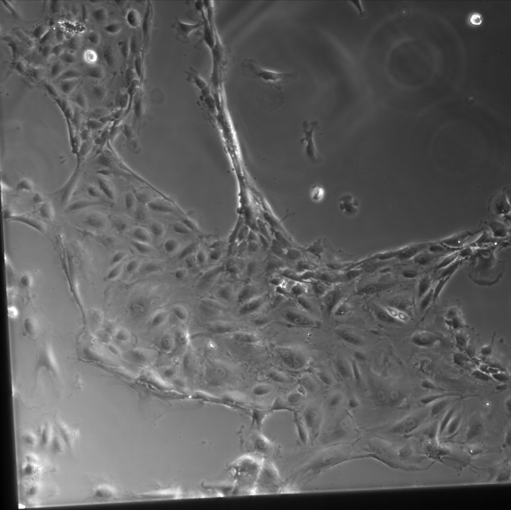
For the first ~94 hours, the cells proliferated as per normal and spread as seen previously on growth rate experiments, seeming to approach dense confluence. However, after about 100 hours, only those cells on the SiO2 membranes began exhibiting strange contractile behavior. The following are the images for each SiO2 location compared to a Tissue Culture Plastic location (all images 10x, scale bar 100µm).
| Time (hr): | SiO2 1-1 | SiO2 1-2 | SiO2 2-1 | SiO2 2-2 | Tissue Culture (TC) Plastic |
| 4.5 | 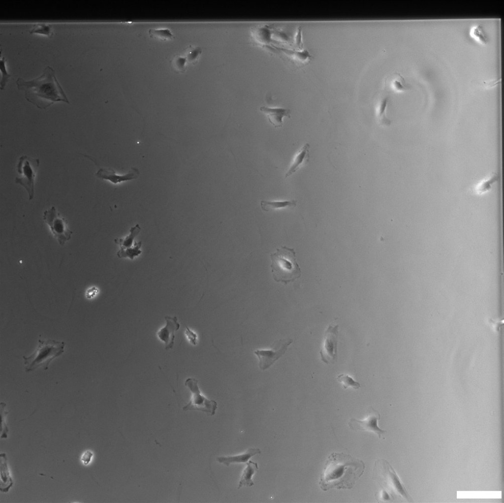 |
 |
 |
 |
 |
| 23 | 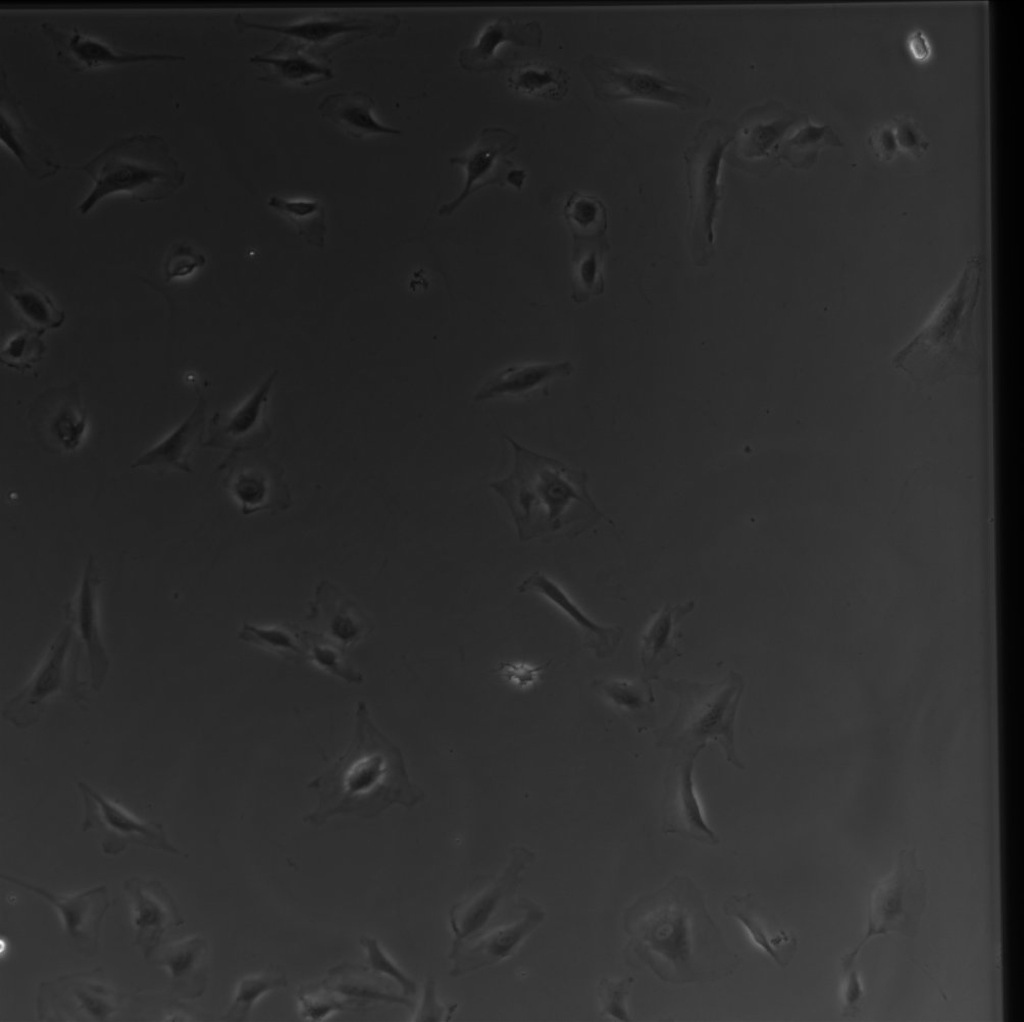 |
 |
 |
 |
 |
| 45 |  |
 |
 |
 |
 |
| 69.5 | 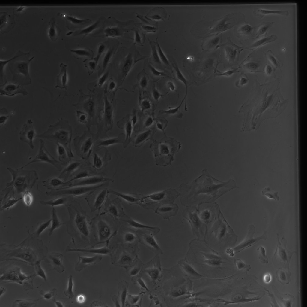 |
 |
 |
 |
 |
| 76.5 | 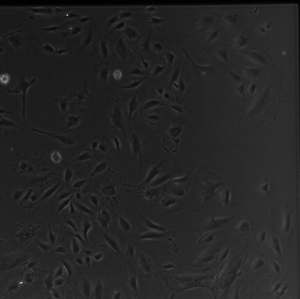 |
 |
 |
 |
 |
| 94 | 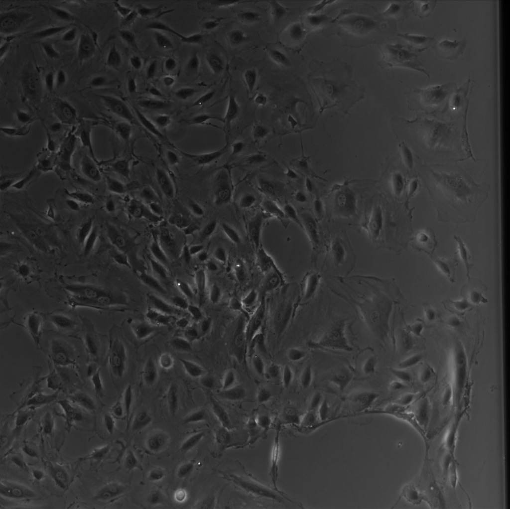 |
 |
 |
 |
 |
| 100 | 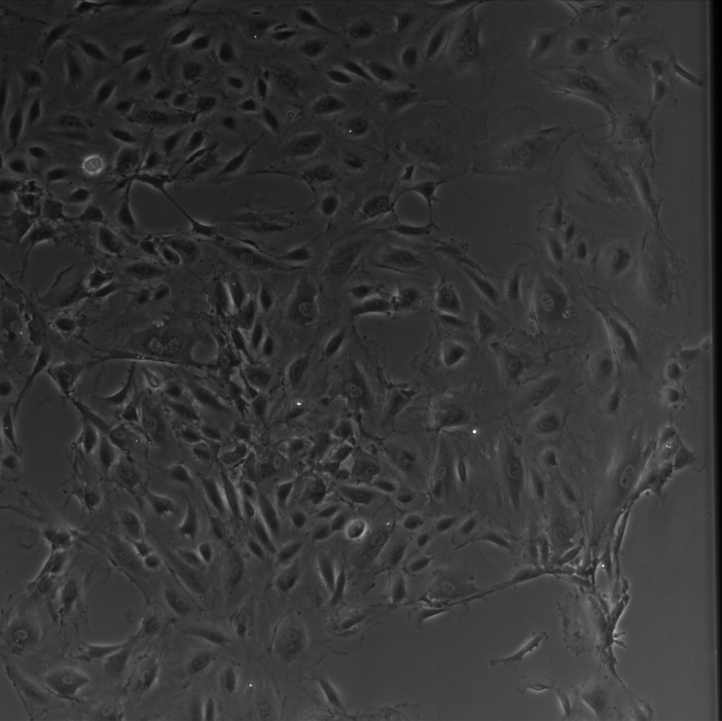 |
 |
 |
 |
 |
| 117.5 |  |
 |
 |
 |
 |
| 125.5 | 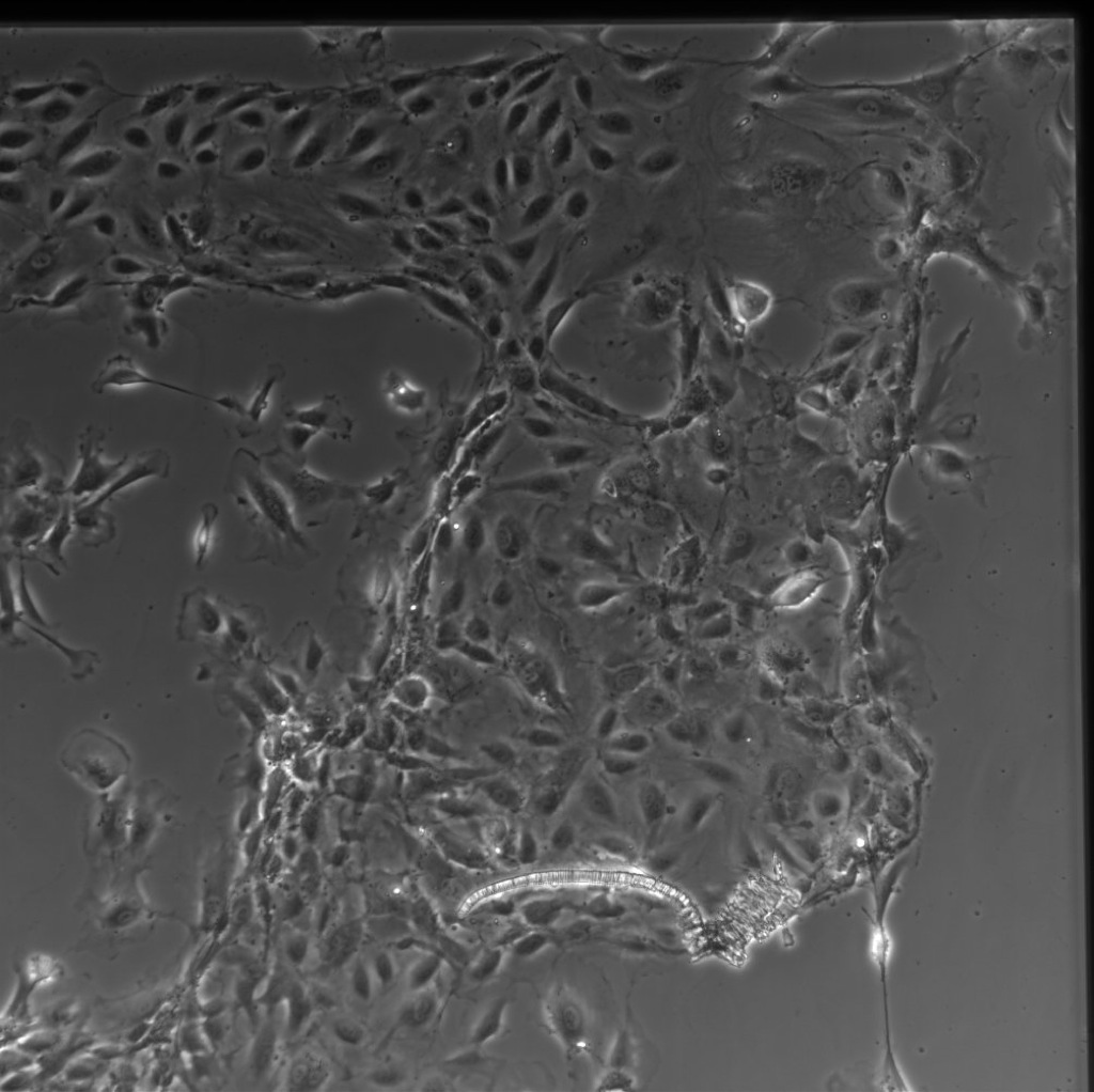 |
 |
 |
 |
 |
| 142 | 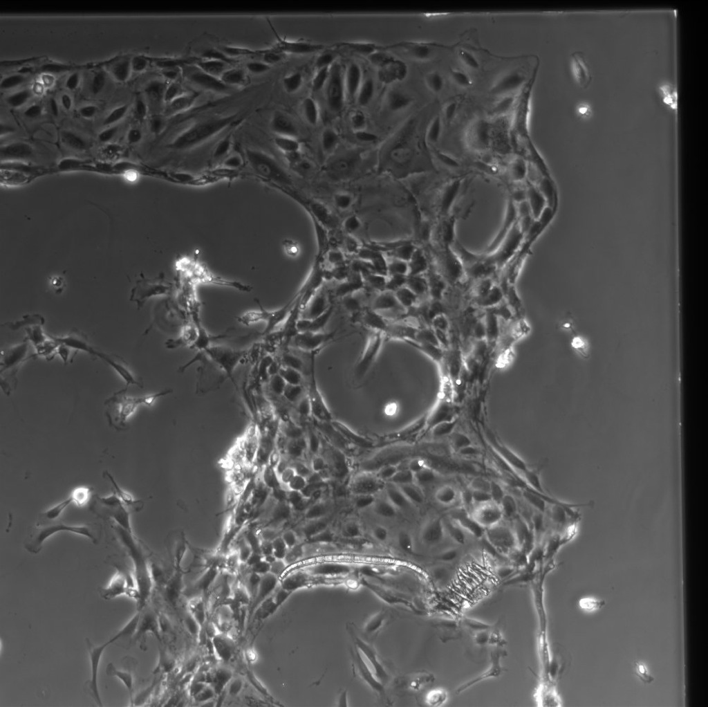 |
 |
 |
 |
 |
| 149 | 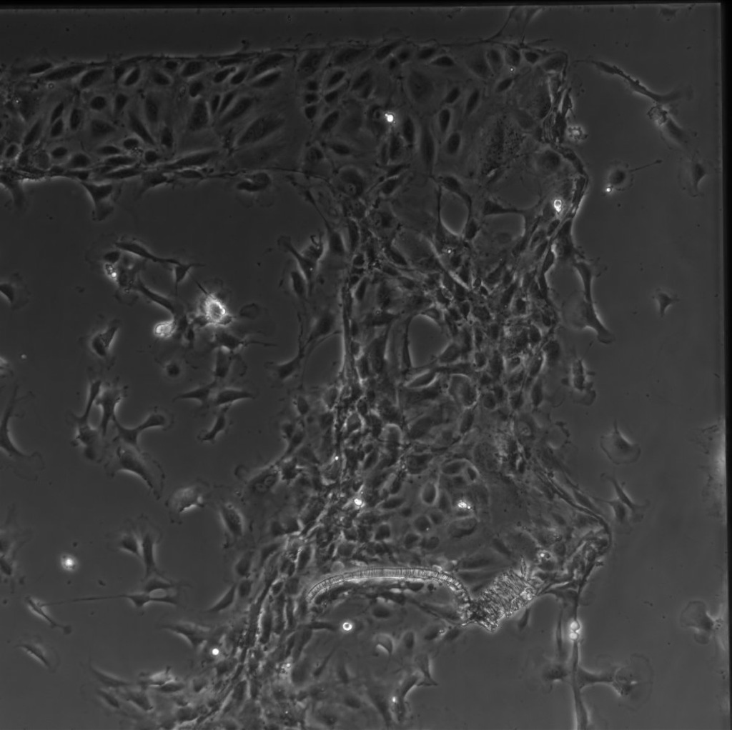 |
 |
 |
 |
 |
| 166.5 | 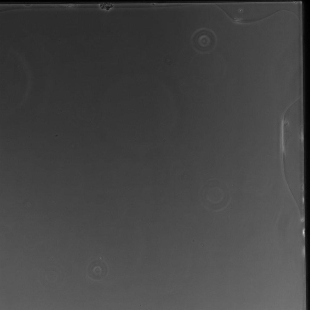 |
 |
 |
 |
 |
| 173 | -membrane broken- | -membrane broken- |  |
 |
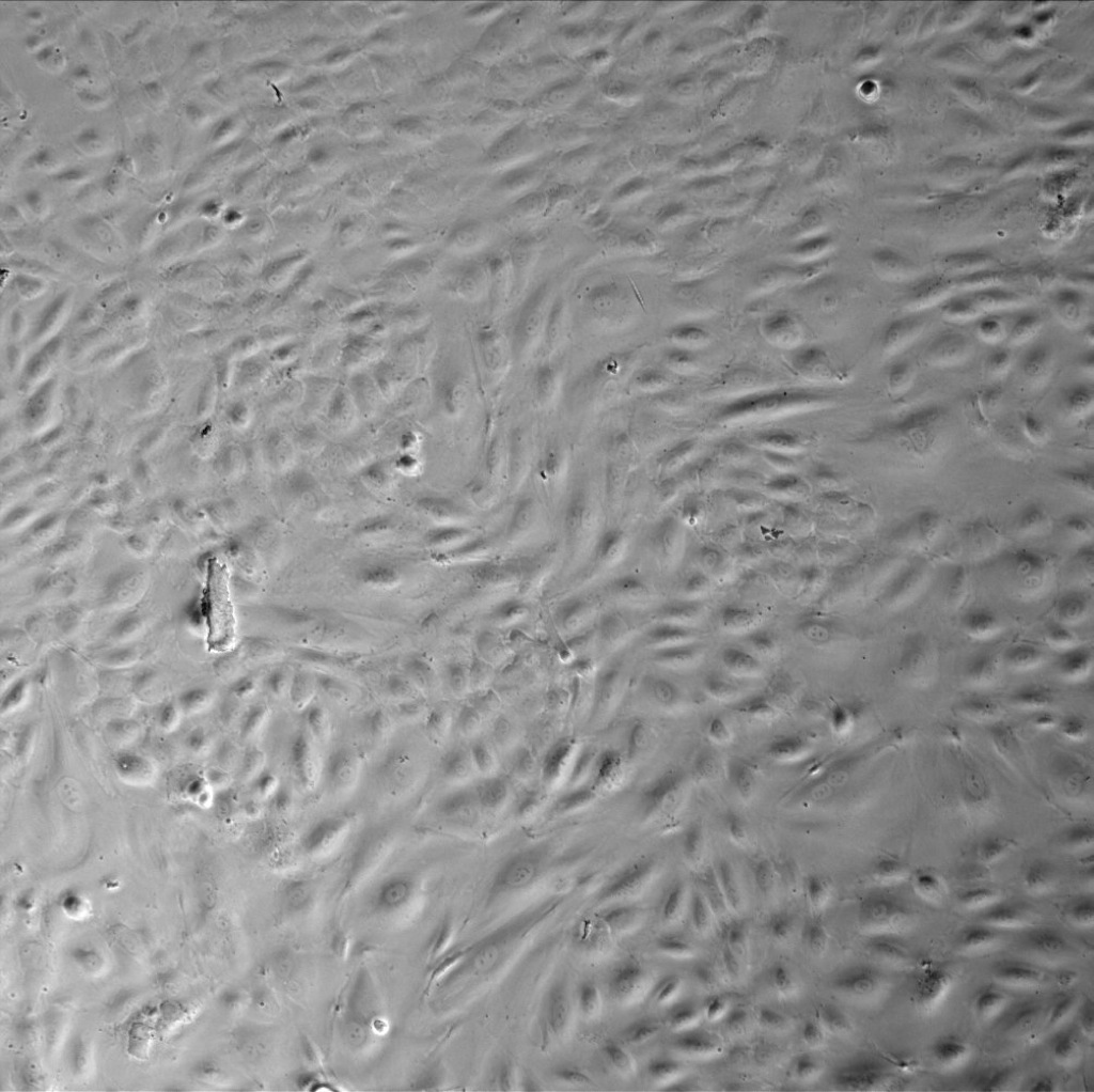 |
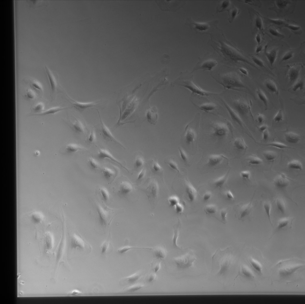
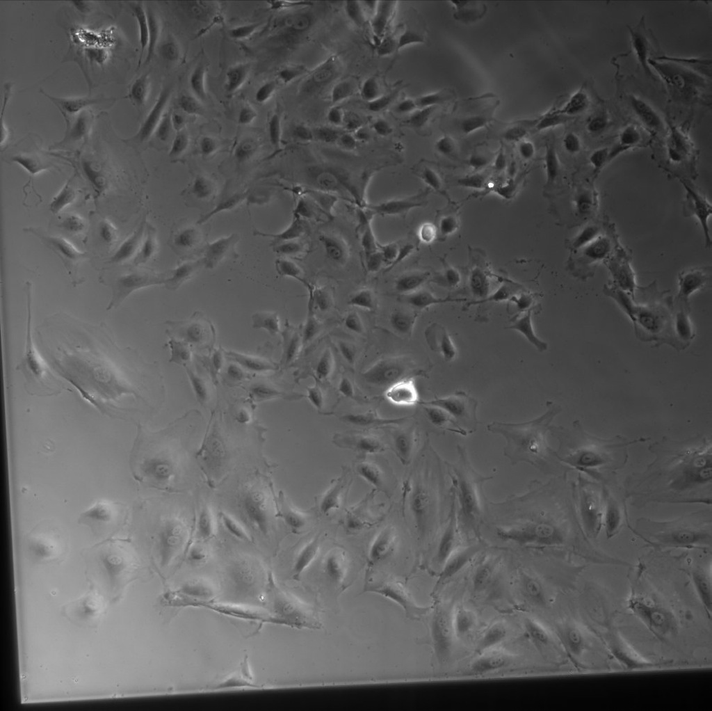
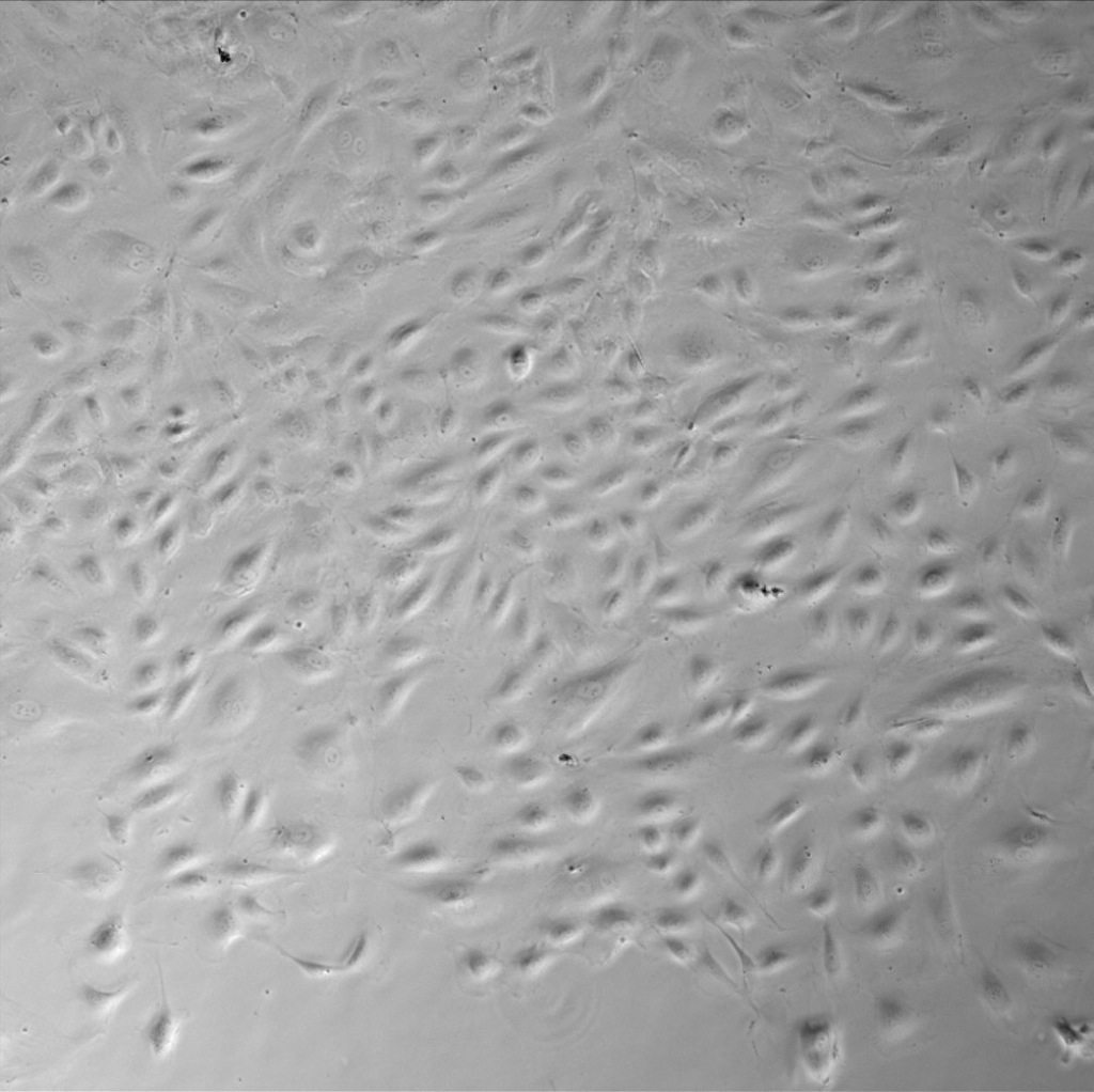
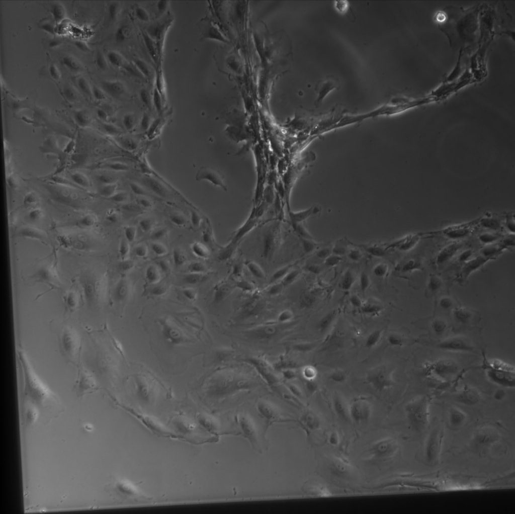
Pulled from this data, here is a comparison of three timepoints of SiO2 (first set of images) vs the same three timepoints at a TC location (second set of images; all at 10x; scale bars are 100µm). The cells on SiO2 are clearly contracting, while the cells on equivalent coating conditions (but on TC plastic) simply proliferate and become confluent:


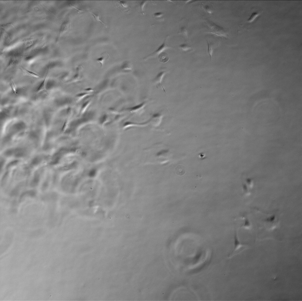
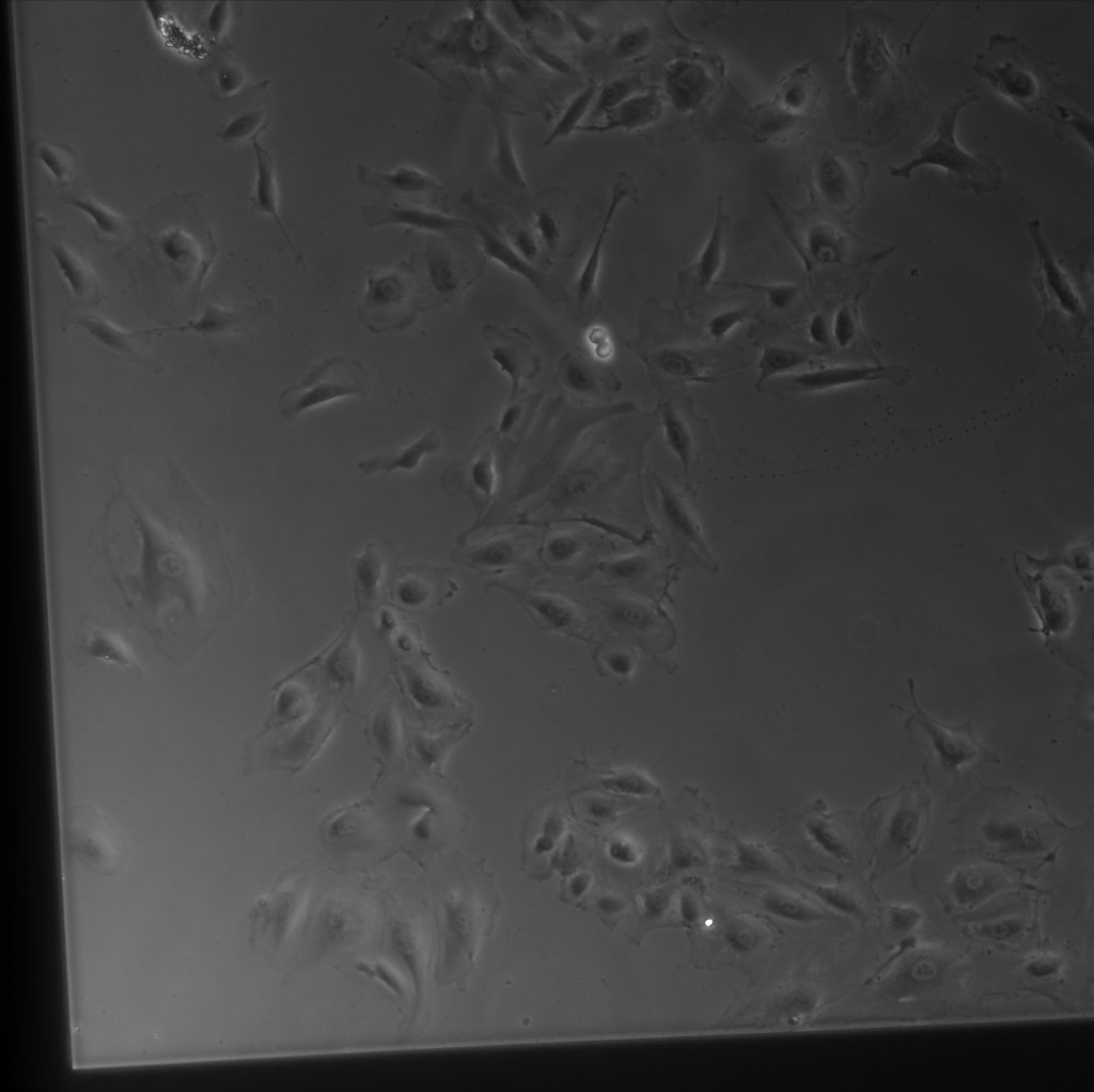
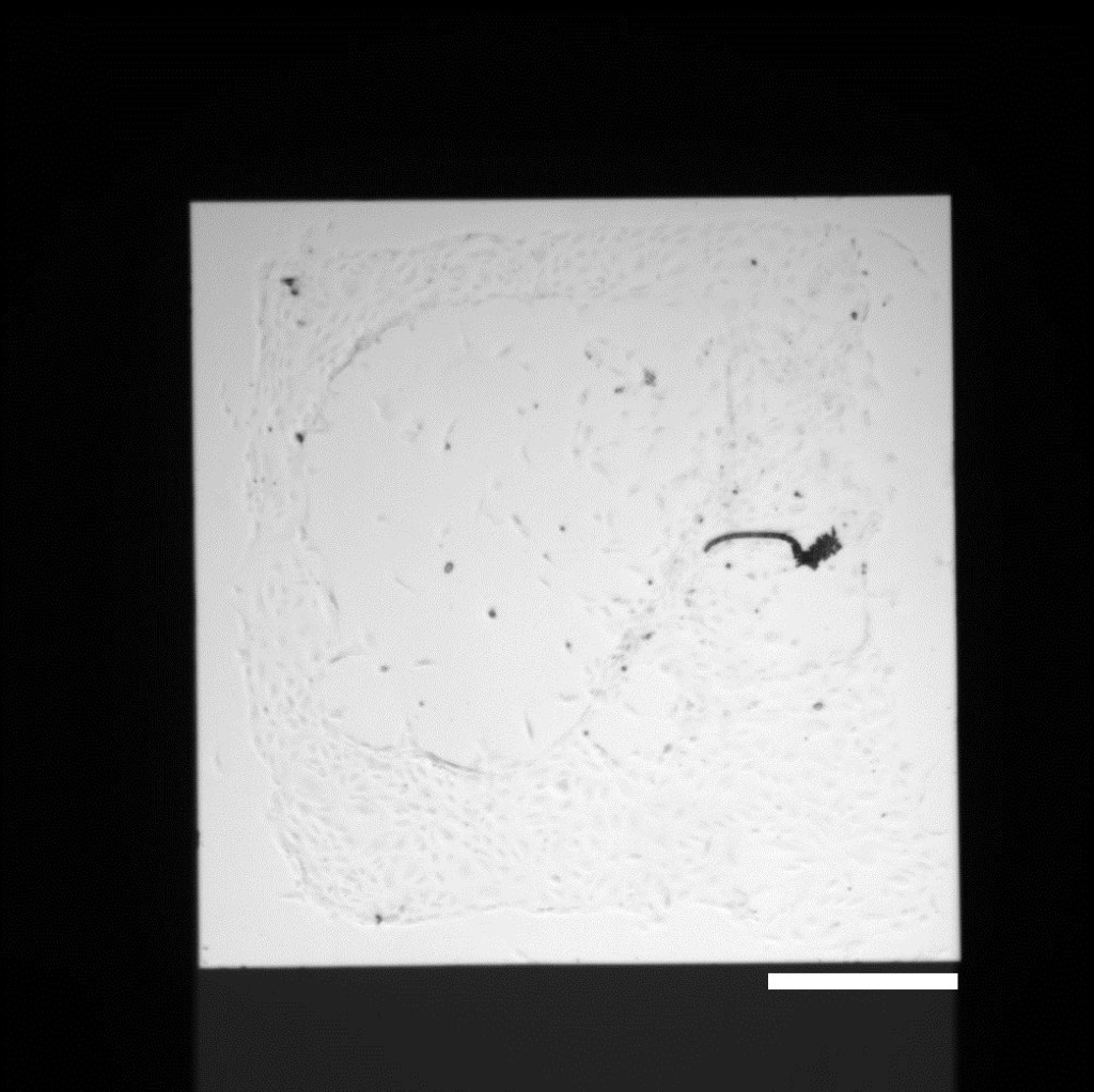
The contraction seems to be at least partially geometrically inspired, as a 4x image (SiO2 1-1 & 1-2 are top-right and bottom-left corner, respectively) at 149 hours shows a clear outline of the square membrane area in the cells’ contraction (scale bar is 500µm):
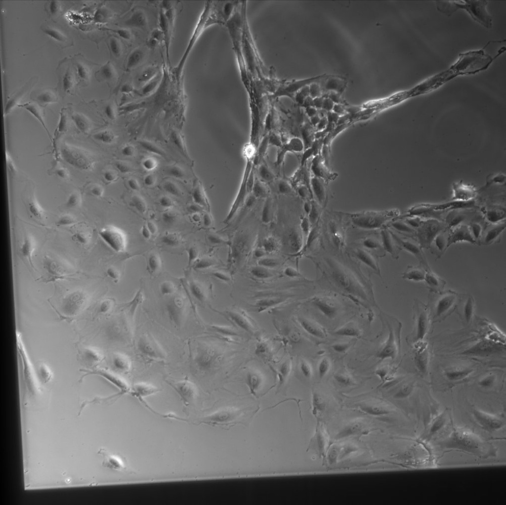
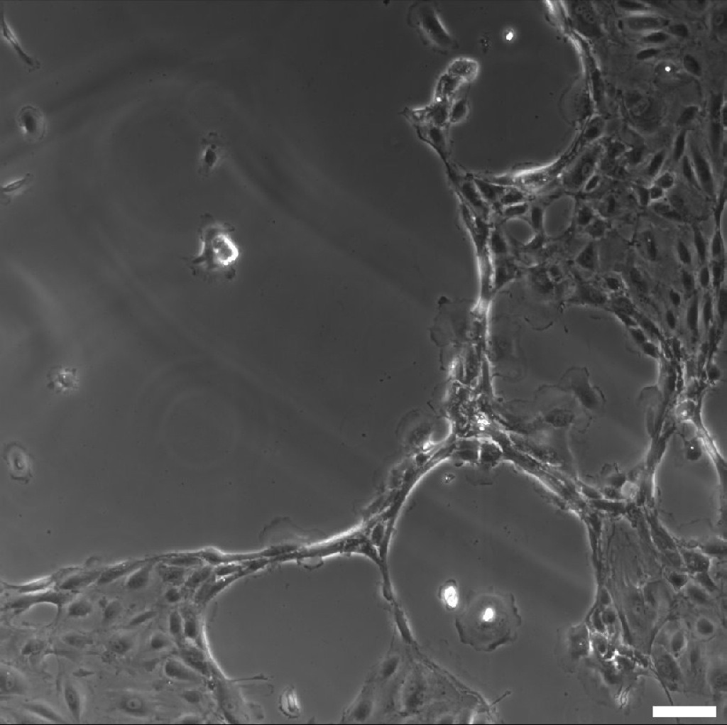
And here is a close-up of the contractile ‘rings’ seen at 166.5 hours (10x, scale bar = 100µm):
I’m not sure what has caused this, but it may be due to the firm geometric constraints. I’ve also received input that it’s possible the membrane has dissolved some in areas, causing some cells to pull away from the surface but remain adhered to neighboring cells. However, it seems that cells can re-crawl into the areas which had previously been pulled away from. This is the first time I’ve observed this phenomena, so it may also be due to the lack of pores (or even the lack of evaporation?).
NRG discussion.
1) Geometrically triggered? Hard to imagine a mechanism because of the hollowing out seen in the middle. Generates strange hypotheses.
2) Keep an eye out for more phenomena like this but don’t follow up.