300nm Thick SiO2 Membrane Soak Test
In this experiment, I soaked membranes (from either wafer 1064 [150W deposition power] or wafer 1065 [100W deposition power]) in different fluids that are standard use in cell culture applications, or which provide a similar situation as could be found in such (cause of pH change, salts, etc.).
Each membrane is made into a single-well CytoVu using 1000µm PDMS gaskets after the chip is clean with IPA. They are then placed in glass petri dishes (one of two sizes, either ~4.5″ diameter [schematic] or ~3.5″ diameter [not shown, used a glass ‘lens’ piece as top]) in the CytoVu-holding slide device, along with a large kimwipe and ~20mL of ultrapure (UHP) water to provide a humidified local environment. See schematic:

One set of membranes (100W and 150W) has 1X PBS placed on top and bottom (30µL and 50µL, respectively) and placed on the benchtop.
The second condition was 0.1M Sodium Bicarbonate, which was created using UHP water and 85mg of the bicarbonate. For these membranes, the same amount of solution was placed on top and bottom as the first, but these were placed in the incubator.
The final condition was DMEM cell culture media with 10% FBS and antibiotics (PenStrep). Again, 30µL on top and 50µL on bottom was used, and these were also placed in the incubator.
A fourth ‘condition’ was simply taking two membranes (one of each deposition power) out of the gel box after the soaking of the others was done, for a control.
The membranes used for the Bicarbonate and DMEM were both UV sterilized for 25 minutes, and also autoclaved before having solution placed down.
All membranes were given one week to soak (the humidification of the petri dish prevented evaporation). After this time, the solutions are pulled off and each membrane is rinsed twice with UHP water, then placed in the oven to dry after aspirating off the liquid.
Images were taken of each of the membranes dry, and non show the wrinkle pattern as was present in the 1000C annealed 120nm thick SiO2 membrane.
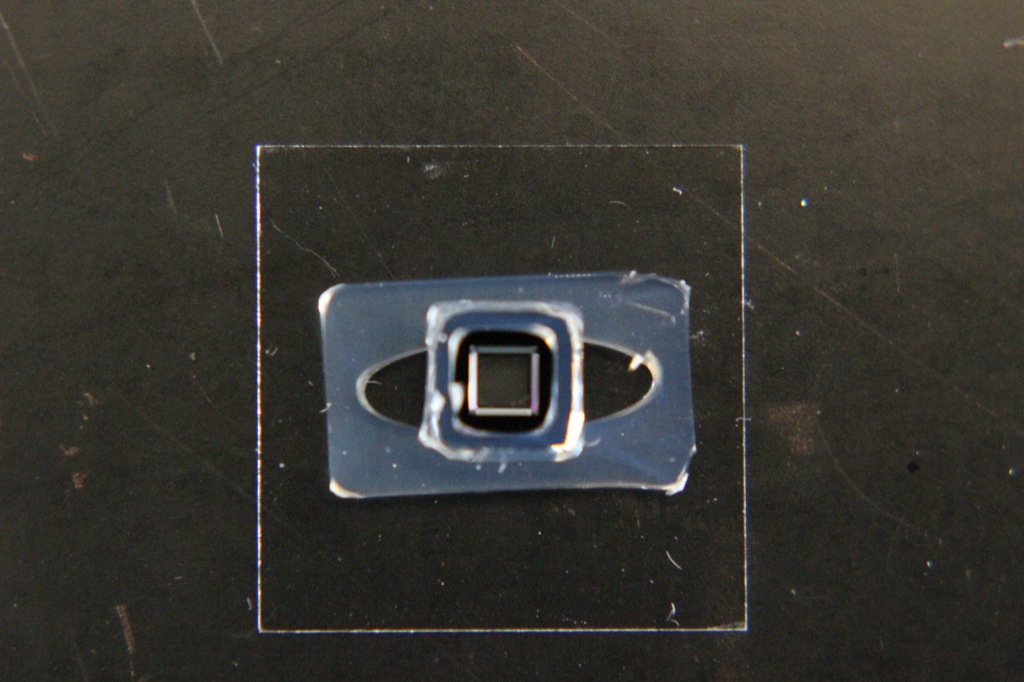
100W 1X PBS:
150W 1X PBS:
100W 0.1M Sodium Bicarbonate:
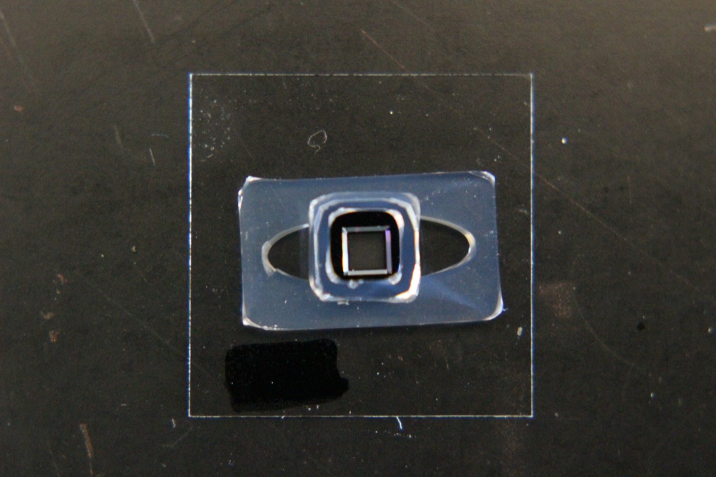
150W 0.1M Sodium Bicarbonate (the line was a hair I didn’t want to remove to prevent damaging the membrane):
100W DMEM w/ 10% FBS & antibiotics:

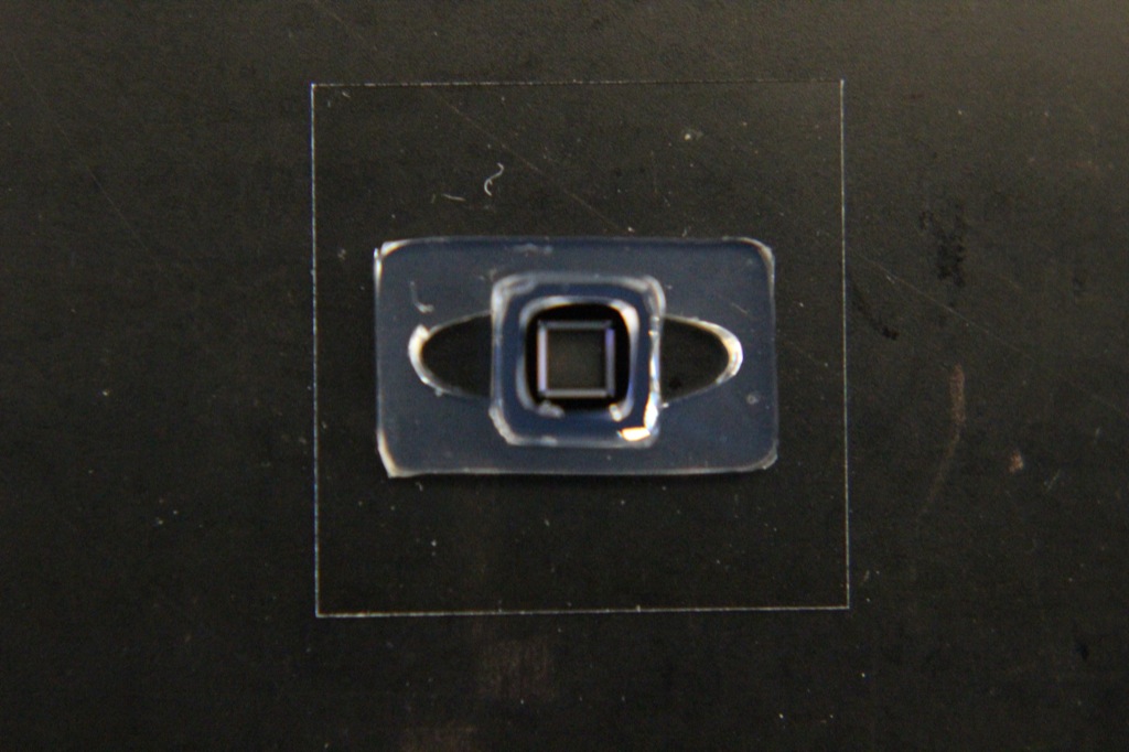
150W DMEM w/ 10% FBS & antibiotics:

100W No Soak (In Gel Box):

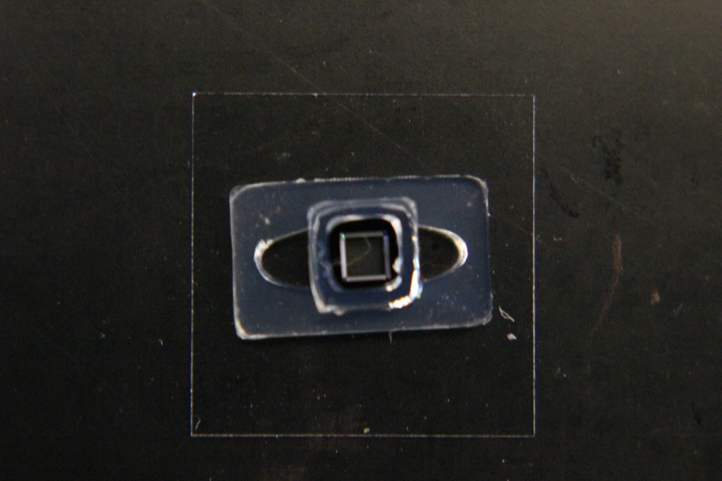
150W No Soak (In Gel Box):

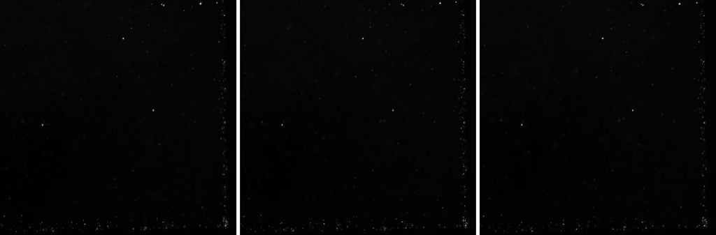
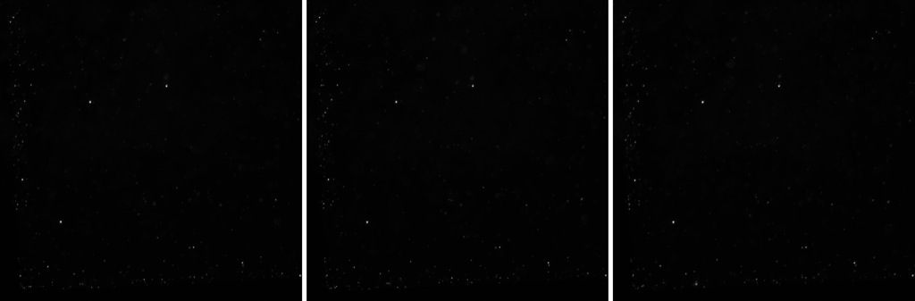
I also performed the bead trial that I had done with the previous experiments. 1µm beads are placed onto each membrane (with UHP water underneath) and allowed to settle over the course of roughly 1.5 hours (Bicarb, DMEM), or 2.5 hours (1X PBS, No Soak). These are then imaged in focused, and 10µm above and below focus to see if any beads are present in differing focal planes still on the membrane (thus indicating the presence of bowing and likely wrinkling in the membrane).
From initial inspection it does seem from the images that there is a good evenness of bead distribution. It is somewhat difficult to analyze, since due to the porosity/liquid on top there can be beads in different focal plans due to floating in solution. However, there does seem to be enough of an indication in each of the images to support a lack of wrinkling/bowing.
The center image is in focus, left is 10µm above and right is 10µm below. Images represent 1/4 of the membrane (roughly 1×1µm). The location of the corner of the membrane is stated for each.
100W 1X PBS (Corner is bottom-right):
150W 1X PBS (Corner is bottom-left):
100W 0.1M Sodium Bicarbonate (Corner is bottom-right):

150W 0.1M Sodium Bicarbonate (Corner is bottom-right):

100W DMEM w/ 10% FBS & antibiotics (Corner is bottom-right):

150W DMEM w/ 10% FBS & antibiotics (Corner is top-left):

100W No Soak (Corner is top-right):

150W No Soak (Corner is top-right):
