MgF2 + Si3N4 Nanomembrane Images
Here are some images of deposited magnesium fluoride films on 50 nanonitride:
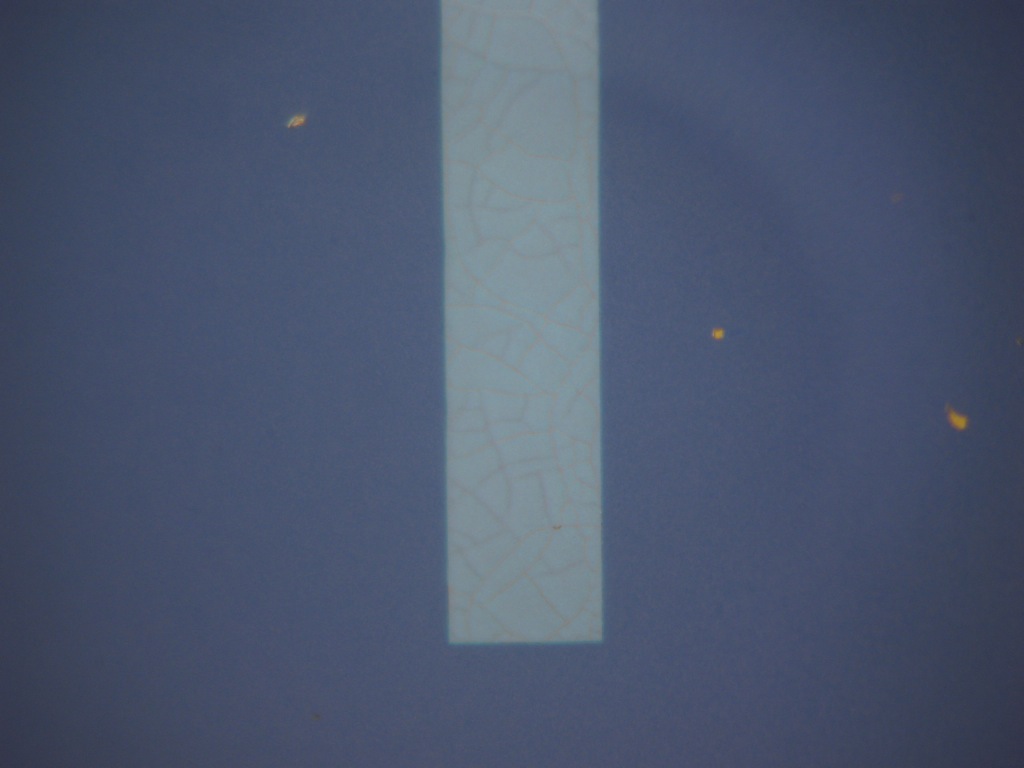
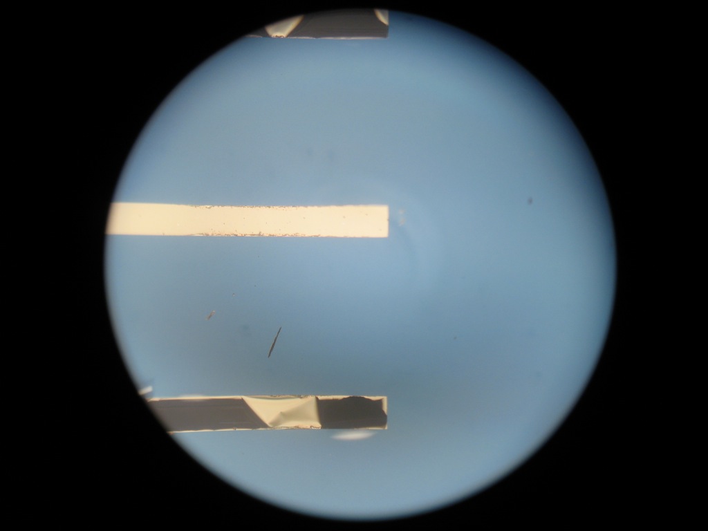
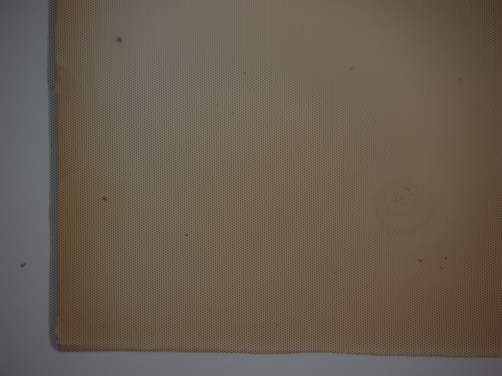



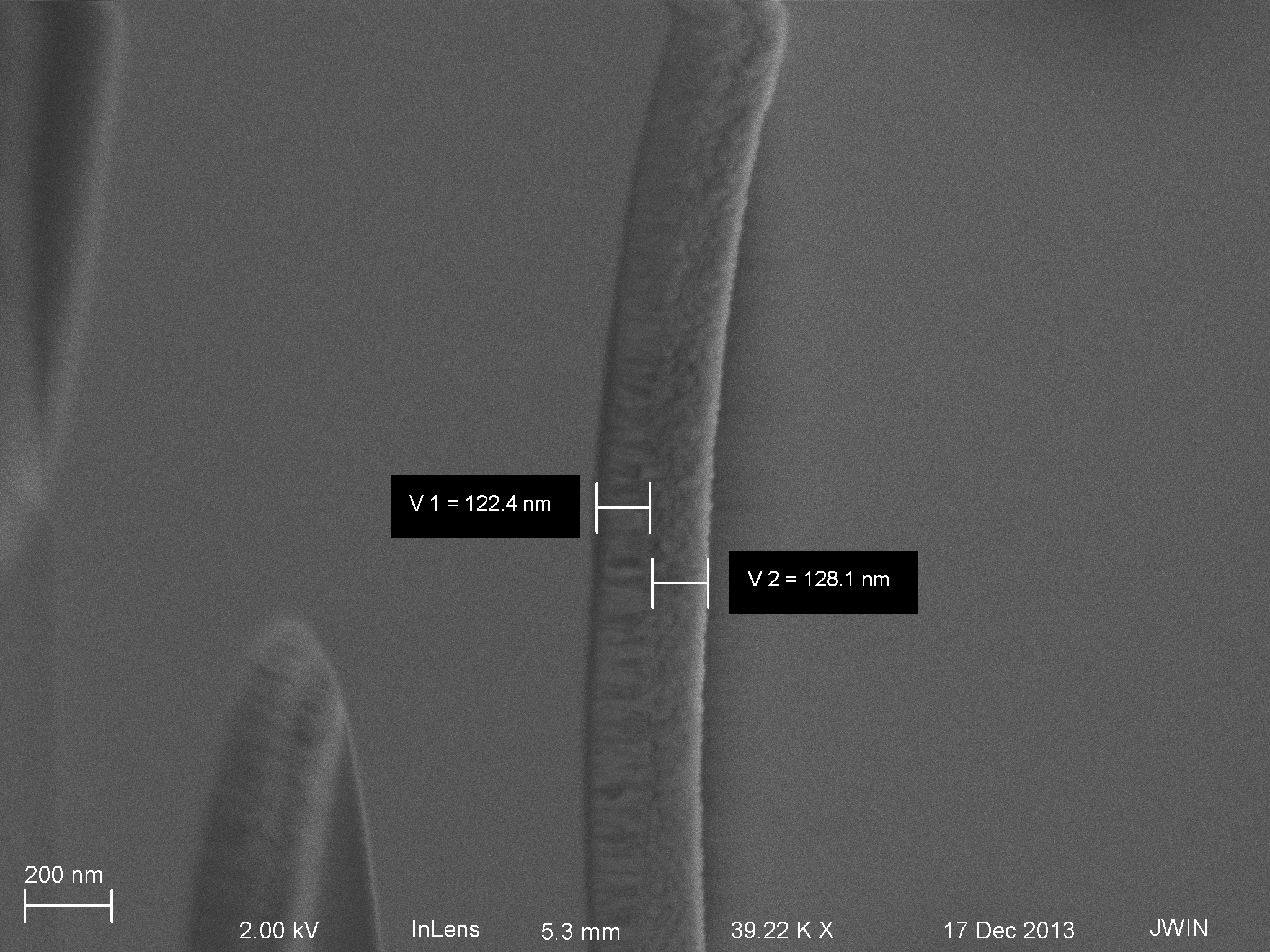
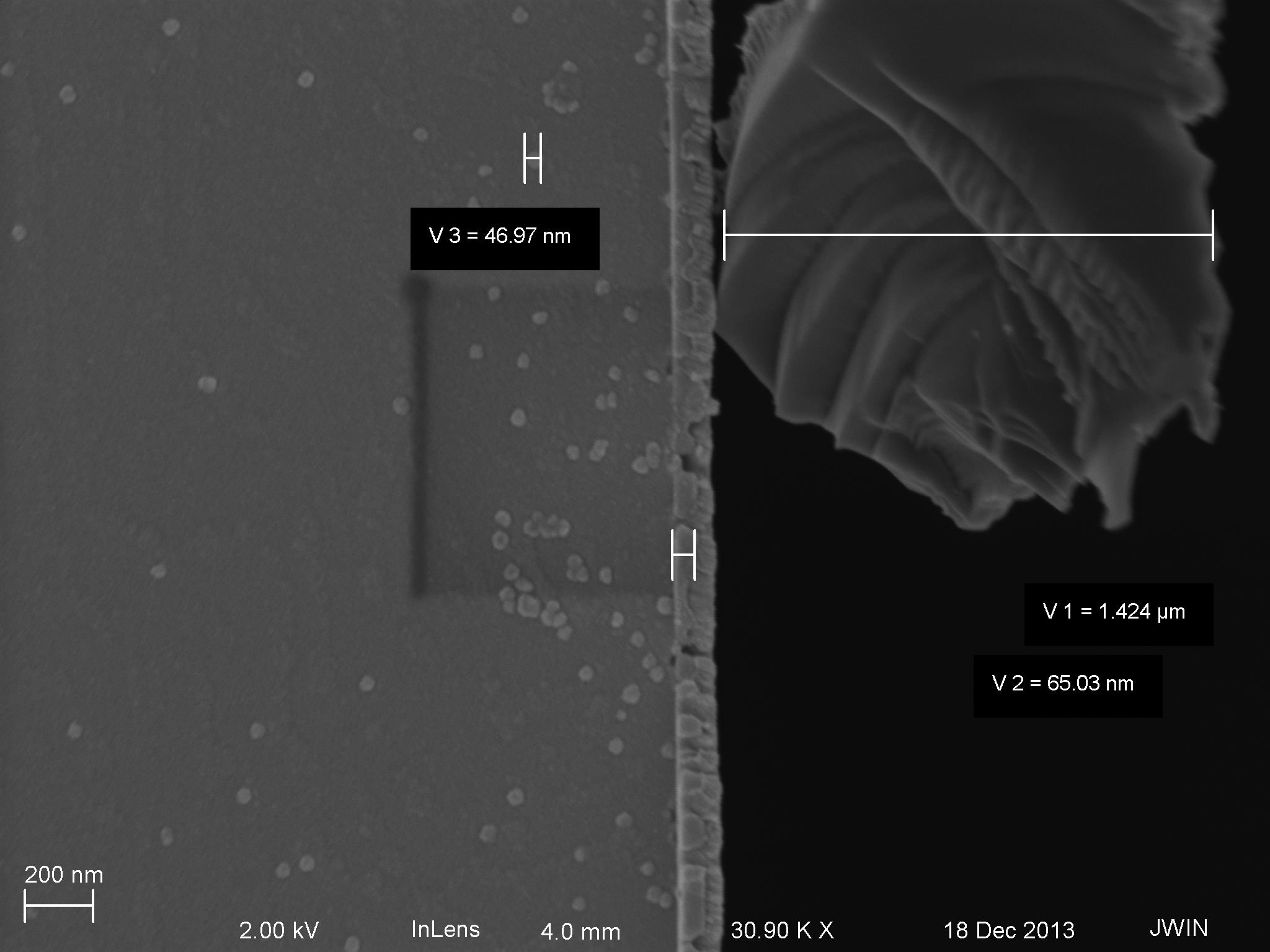

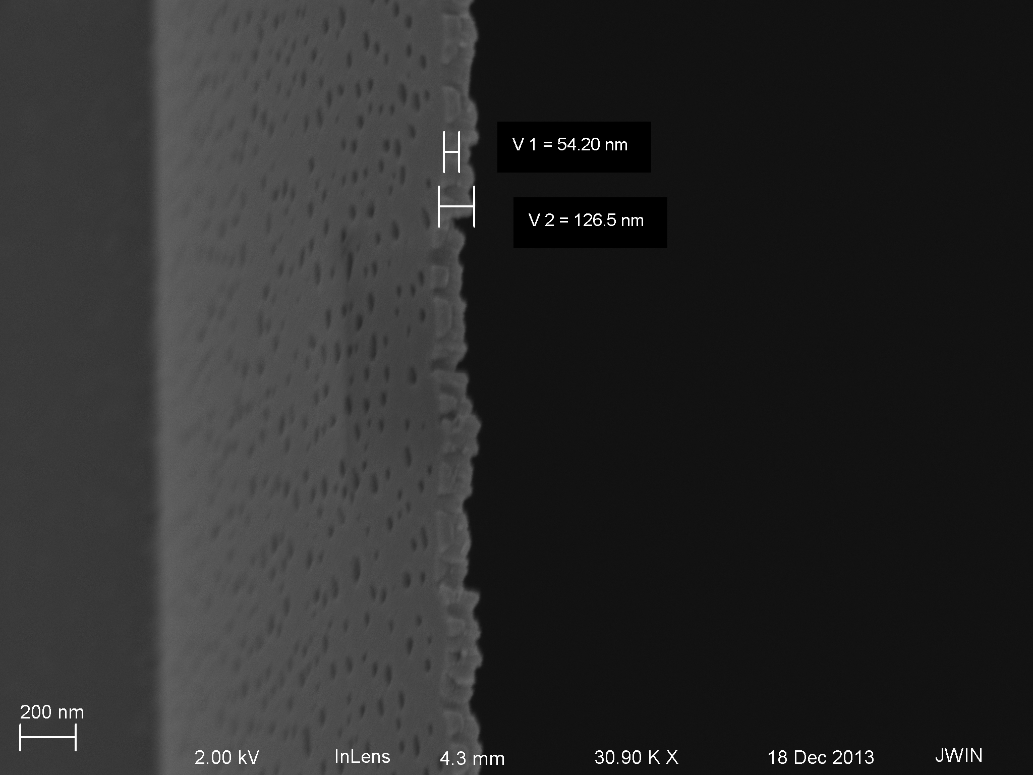

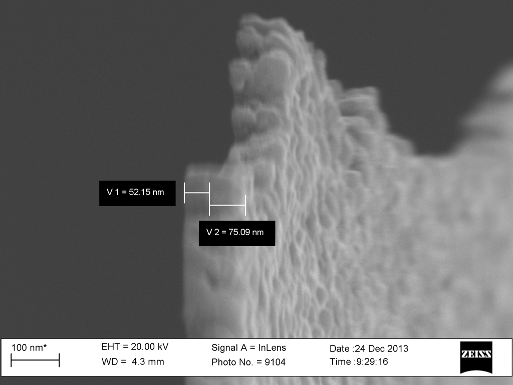


Here are some images of deposited magnesium fluoride films on 50 nanonitride:
Hi all, We have already showed in our earlier [ post ] that our shear-free chemotaxis system can indeed elicit chemotaxis, suggesting that we have successfully translated a gradient through the membrane to the shear-free side of the system where the cells reside. However, it is still nice to show via fluorescence how the translated gradient looks…
For the first time, we did time-lapse phase microscopy (1 frame / 5 seconds) looking at endothelial cells through the ITO in the static BBB device (device A bottom left well) and measuring the TEER concurrently. Here’s the video, just shy of 15 minutes of recording. (reporting a low resistance: 5-10 ohms, shorted). TEER Test…
Last week, five wafers were produced – three had a double 3500 A wet oxide growth/strip and two had a single 3500 A wet oxide growth/strip. From Jess’ AFM work, an as-is wafer showed a high density of 5-7 nm particles on the wafer surface. With a double oxide growth/strip, these particles do not show…
Using the techniques described in my previous post: TEM Tomography of NPN and npMgF2, we have recreated some tomograms that have contour (position) and height (depth) information contained in them. Our goal now is to express that height information in the form of a surface, or volume. Segmentation Having some evidence that…
We received W648 from the production team this week. Upon inspection of this wafer, is has a higher density of pinholes than we have been seeing lately (>3 pinholes/slit compared to 0 or 1/slit) and seems to show a return of contamination, shown below. This contamination is visible over the entire wafer, but is easiest…
[presentation] HUVEC migration and fibronectin