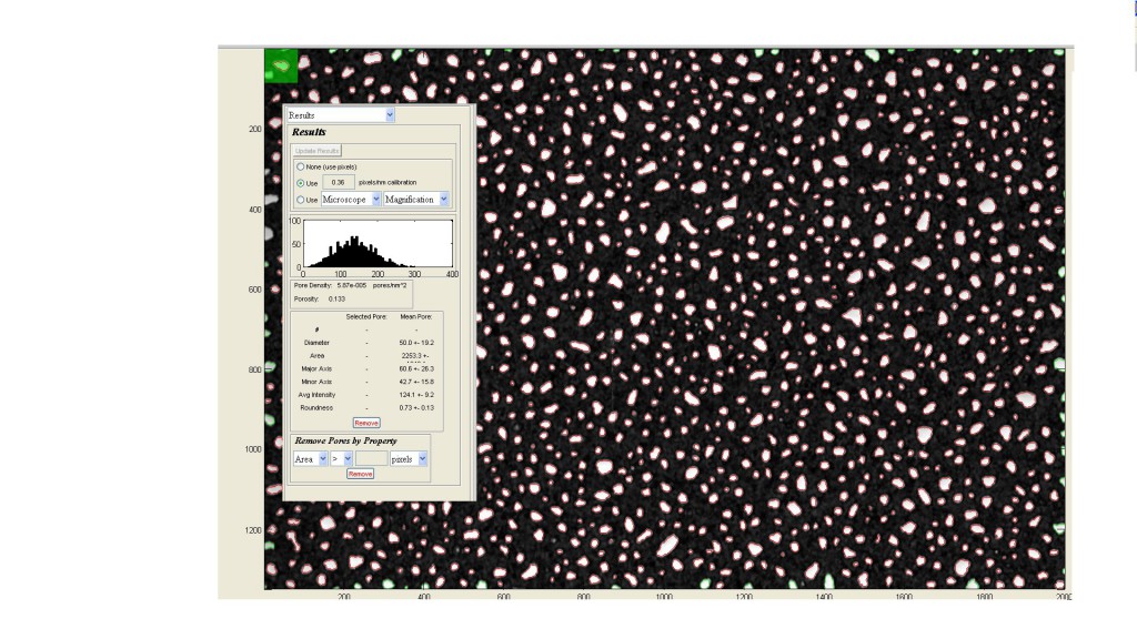
I have tinkered with the etch recipe some more, adding Argon to help prevent polymer formation (adding 5% O2 was not enough). The current recipe is 200 mTorr (100 mTorr Ar, 5 mTorr O2, 95 mTorr CHF3) at 120-125 W (6-12 W reflected). The plasma color is initially rosy (Argon), settling into a light purple after a few seconds (Light blue from CHF3 and O2 mix). After 140 seconds of etch using this recipe, the coated (250C, 1-3 A/s, 50nm) MgF2 films remain, while the backside is fragmented and discontinuous.
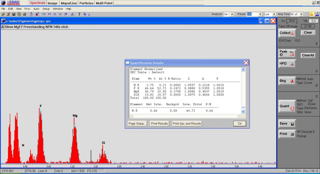
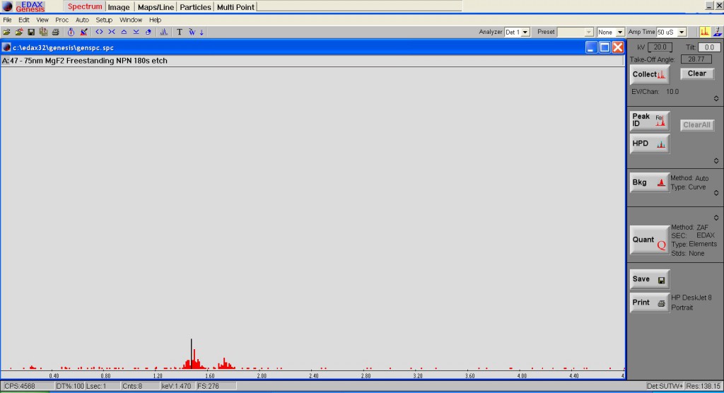
The true composition of these films has yet to be confirmed. Using EDAX on our SEM, I was able to find a decrease in the relative Silicon composition of the etched film, though some signal remains. I hope to get an XPS reading soon to confirm this result.
In the discontinuous film state, the etched free-standing membranes have survived for 48+ hours. The burst pressure for these films (SEPCON format) were 1.5 and 1 psi, much weaker than what we are used to having from nitride alone (thermal processing after deposition to densify the films may help in the future).
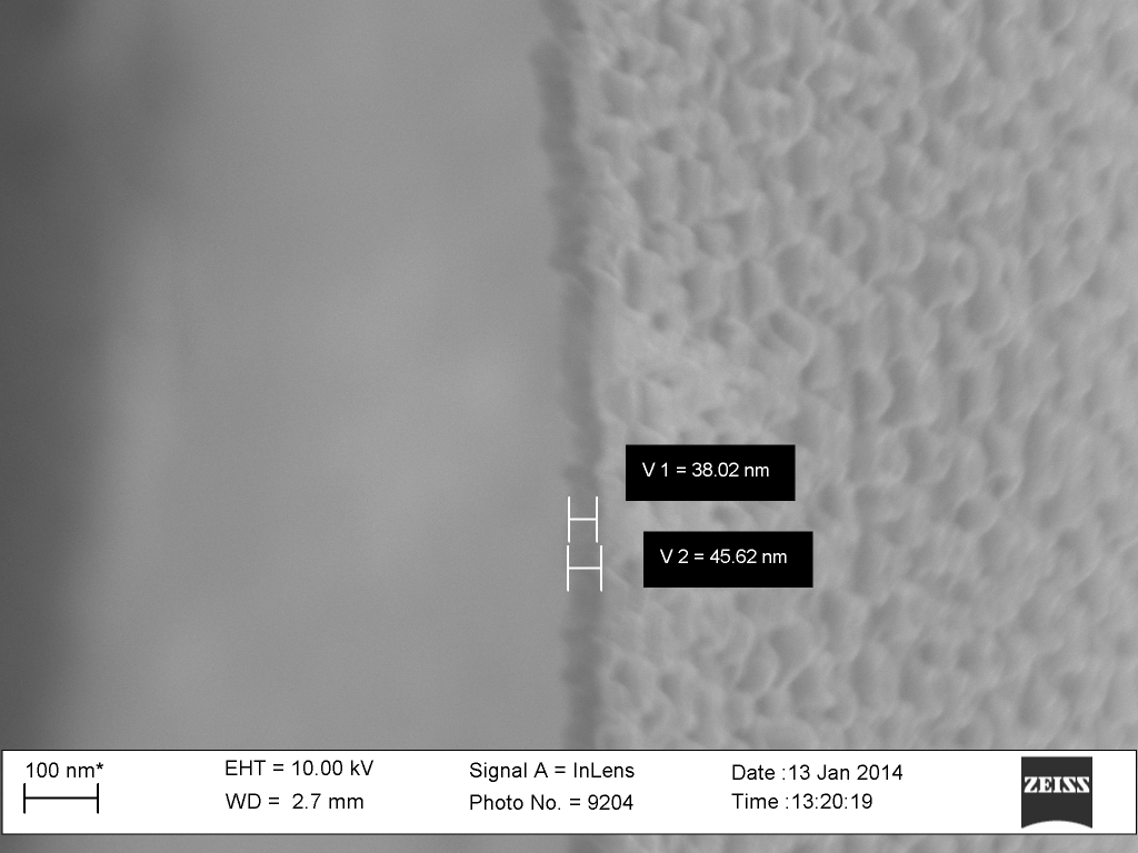
Step height on 50nm MgF2 film (kapton tape occludes deposition surface)
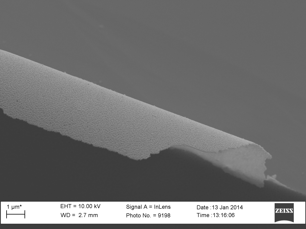
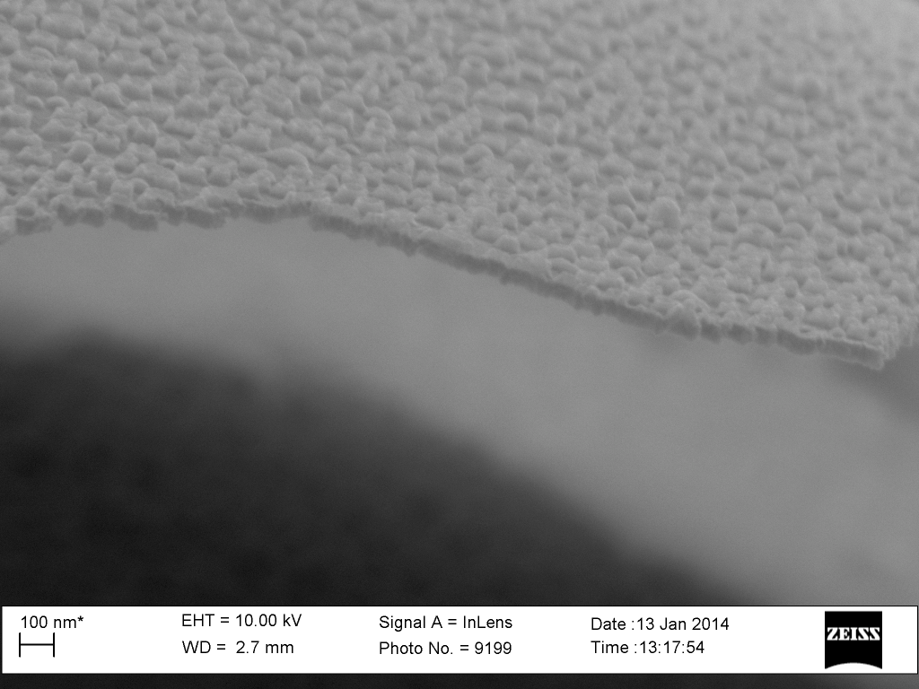
Sample 40, 50nm MgF2, 140s Ar+CHF3+O2 etch, curling up from freestanding region. The dark region is the bulk. The top face would be where the silicon nitride has been etched (backside)Sample 40, 50nm MgF2, 140s Ar+CHF3+O2 etch, curling up from freestanding region. The dark region is the bulk. The top face would be where the silicon nitride has been etched (backside). The film appears discontinuous, with little volcano structures remaining. Ideally, we would have none of these structures remaining with a more complete etch.Is the etched Film Pure MgF2?Or is the remaining film scummed up with Si3N4 volcano structures around the pore?
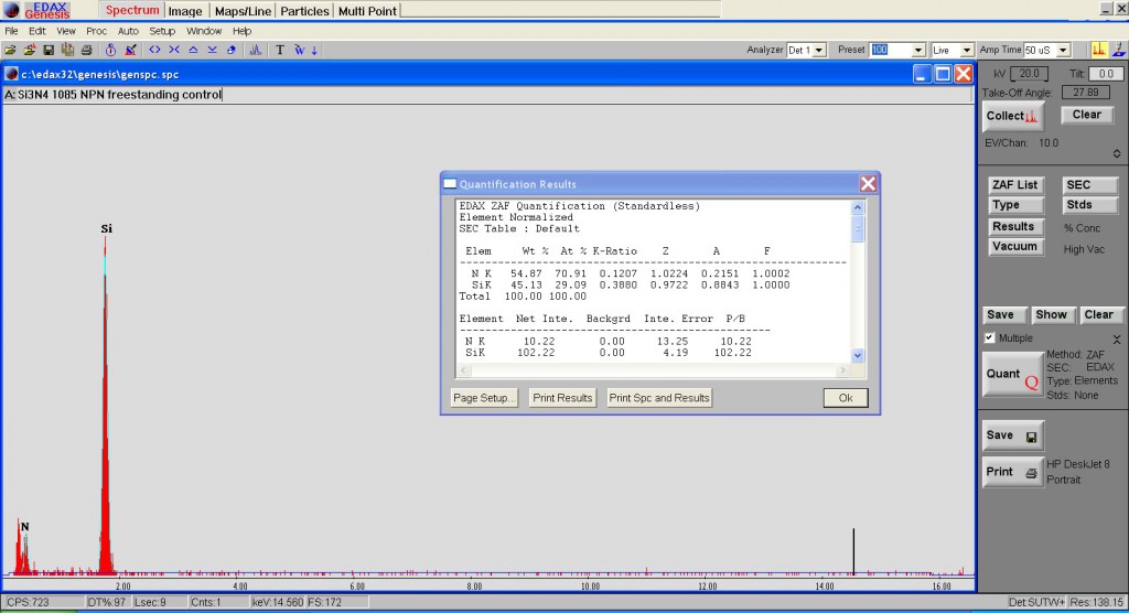
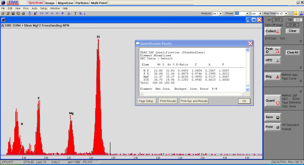
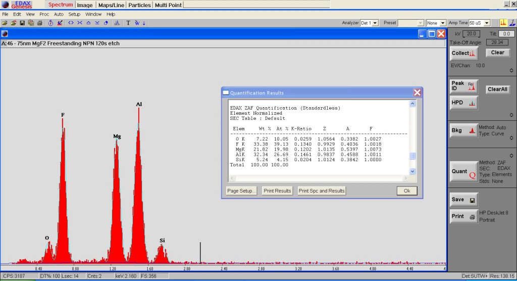
Control 50nm NPN EDAX measurement. Pure Si3N4 is expected.50nm NPN with 50nm of MgF2. This film has not been etched.140s Etched MgF2 membrane. Little silicon remains in the signal (85% MgF2). The larger peaks on the left are the Carbon background.I tried measuring another etched film with a different holder (Al). Again, the silicon signal remains. This sample had 120s etch (compared to 140s).This sample had 180s of etch, but no signal is coming out of the membrane? Clearly a freestanding region under SEM, and a measurement on the sidewalls of the chip confirm the EDAX detector’s functioning.
Sample 40, 50nm MgF2, 140s Ar+CHF3+O2 etch, curling up from freestanding region. Film thickness of MgF2 film is preserved.
Sample 40, 50nm MgF2, 140s Ar+CHF3+O2 etch, curling up from freestanding region. Crossection shows the thickness of the MgF2 film remains the same.Sample 40, 50nm MgF2, 140s Ar+CHF3+O2 etch, curling down in freestanding region. MgF2 surface appears much smoother, with through pores apparent.
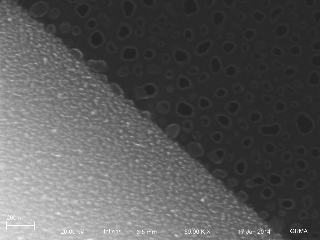
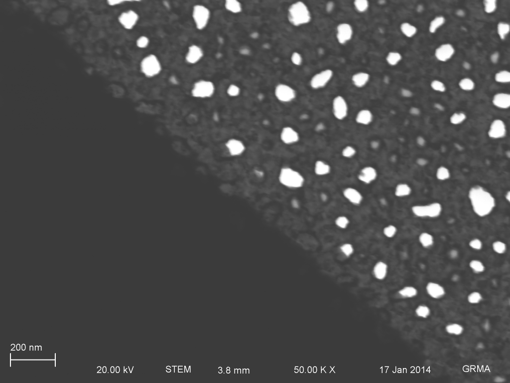
Sample 47, 180s etch SEM mode.Sample 47, 180s etch, TEM mode.
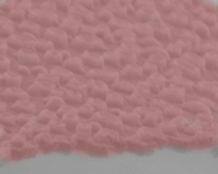
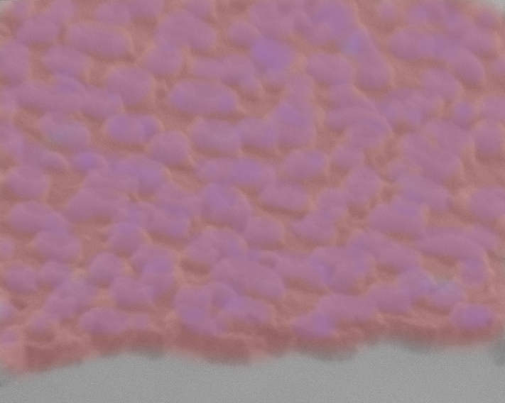
47 – Pore Histogram
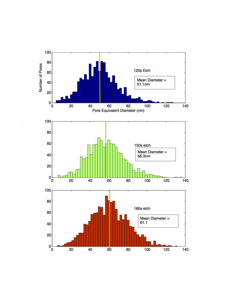
Pore Histograms from samples 45, 46, and 47. Sample 45= 150s, 46=120s, 47=180s. Etching longer appears to blow out the pores somewhat, though the resistance is fairly high for 60s of additional etch time.
Last summer, Tejas and I spent a few days beating our heads against the wall because I was trying to develop a mathematical model of particle capture. Specifically, I was trying to understand what amount of transmembrane pressure we would need to hold a particle on (or in, as we are a politically correct, non-discrimination…
To identify how much of an effect the membrane has on particular simulations, we originally came up with the idea of using a phenomenological effective diffusion to view the membrane impact (see this post). Here is the most up to date version of the figure, which was slotted for use in the simulated diffusion paper:…
After reading and discussing a paper in journal club that talked about cells having a “mechanical memory” (found here “Mechanical memory and dosing influence stem cell fate“), we decided to replicate our previous YAP/TAZ experiments with passage one HUVECs direct from Life Technologies as to avoid making our cells “remember” that they had ever been plated…
Kevin Healy is a Phase I and Phase II awardee of the NIH Tissue Chip initiative. The goal of this initiative is to create ‘mini-organs’ ‘organs-on-a-chip’ ’tissue chips’ or ‘microphysiological systems’ (MPS) for higher throughput and more reliable drug discovery. The effort is also motivated by the failure of recent animal models to predict drug…
In this post, I’m going to discuss the results of the different PEG molecular on silane samples. We used same condition to make 4 chips for each sample. And we treat them with BSA, IgG, and PBS to rest the adsorption. We are using the SEM, contact angle and XPS as the testing method. The way we…
Hi all, a quick update in case you want to know what we are up too. This summer it will be just me at the McGrath lab; the main goal is to produce some preliminary data with an eye on submitting an NSF grant in the fall. The topic is nano-biosensing, using Henry’s shear-free…