SEM of Nanoparticles and NPN
Below are various SEMs of NPN membranes. (All samples were coated with metal by using the Denton Ag sputterer for 120 s) These were used samples that have been sitting out in the lab for weeks. I don’t remember the exact details of the processing. I believe they were filters we used to test the possibility of using our filters under constant flow (as generated by a syringe pump). This tells me we might be able to learn a good deal by looking at samples after filtrations.
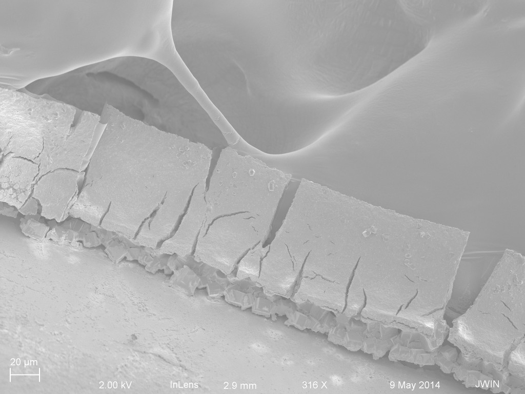
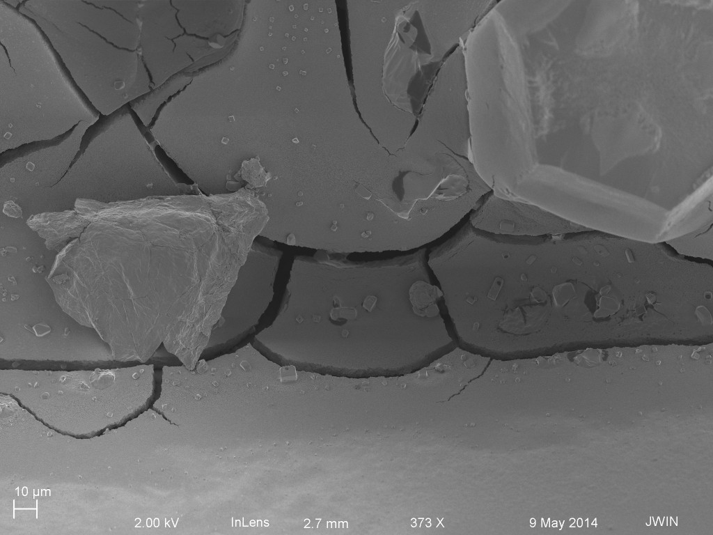
The first image is the backside of a trench. The bottom left corner is wafer, the rectangular strip is the backside of a window, and the upper right corner is the carbon tape from the sample holder. The backside of the membrane has a very thick layer of caked NPs. What you are seeing becomes clearer as I increase magnification.
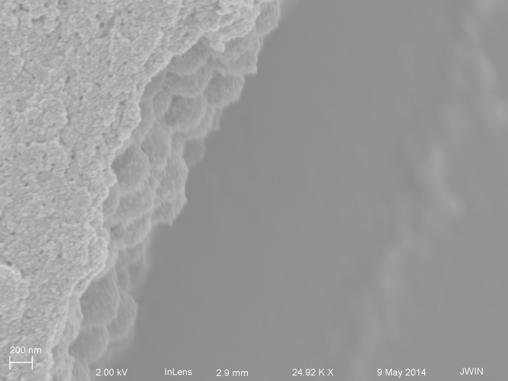
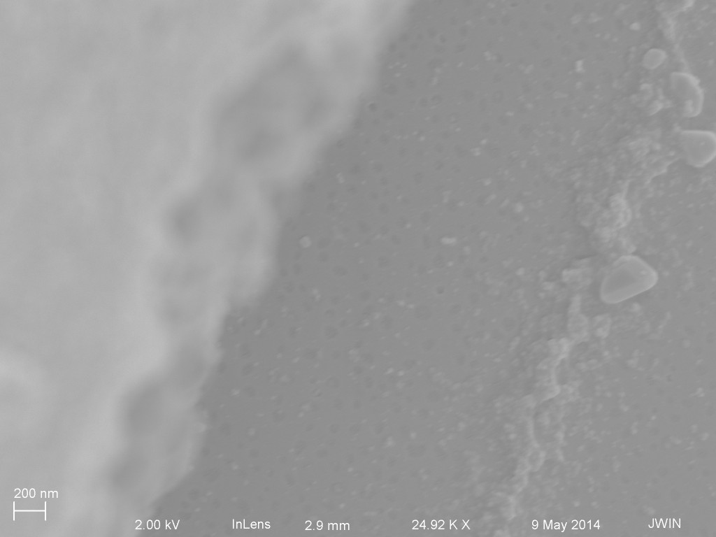
This is looking at one of the cracks in the film you saw above. The left is the “cake” layer of NPs. Just a massive collection of NPs. On the right is the membrane, out of focus at a lower level than the cake layer.
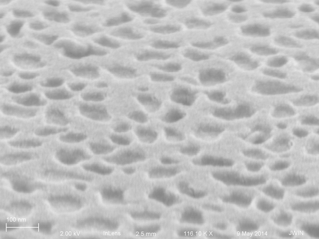
Here we focus in on the membrane below.
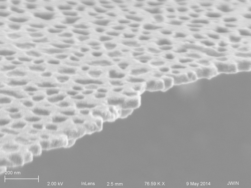
The micrograph below is showing the top side of the membrane we just saw above. So there is no cake layer. We don’t know for sure if the particles passed through the membrane because typically these filters broke during the test and would have NPs on both sides because of that.
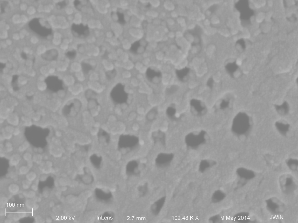
Another topside shot with the tear in the membrane at the bottom.

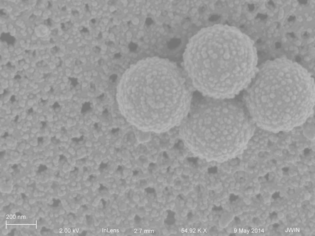
At some point I was doing tests with 500 nm NPs mixed with 20 nm NPs. I assume that’s what we are seeing in the next micrograph.
The next micrographs are of a filter from 1081, but using 100 nm NPs. Again, we see a massive cake layer on the backside of the membrane.
Increased mag to look at one of the cracks.

Inside the crack, still more NPs. I can’t say for sure if we can see the membrane.

This is the top side. The membrane is torn diagonally. On the left is the caked 100 nm NPs. On the right is the membrane.
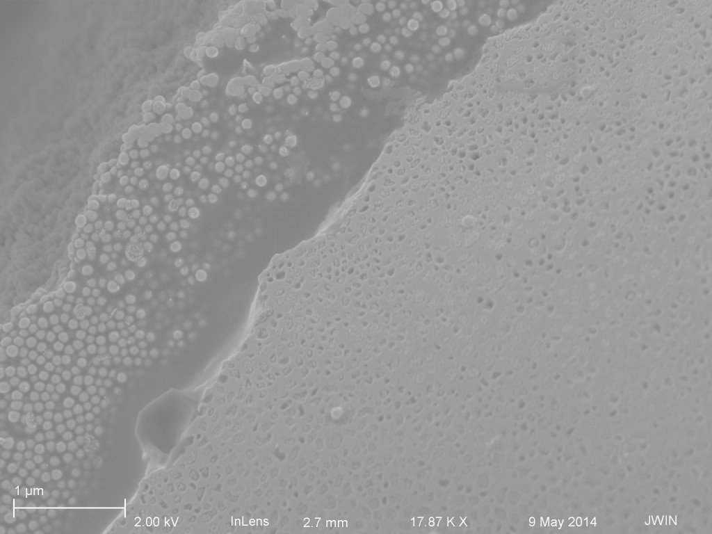
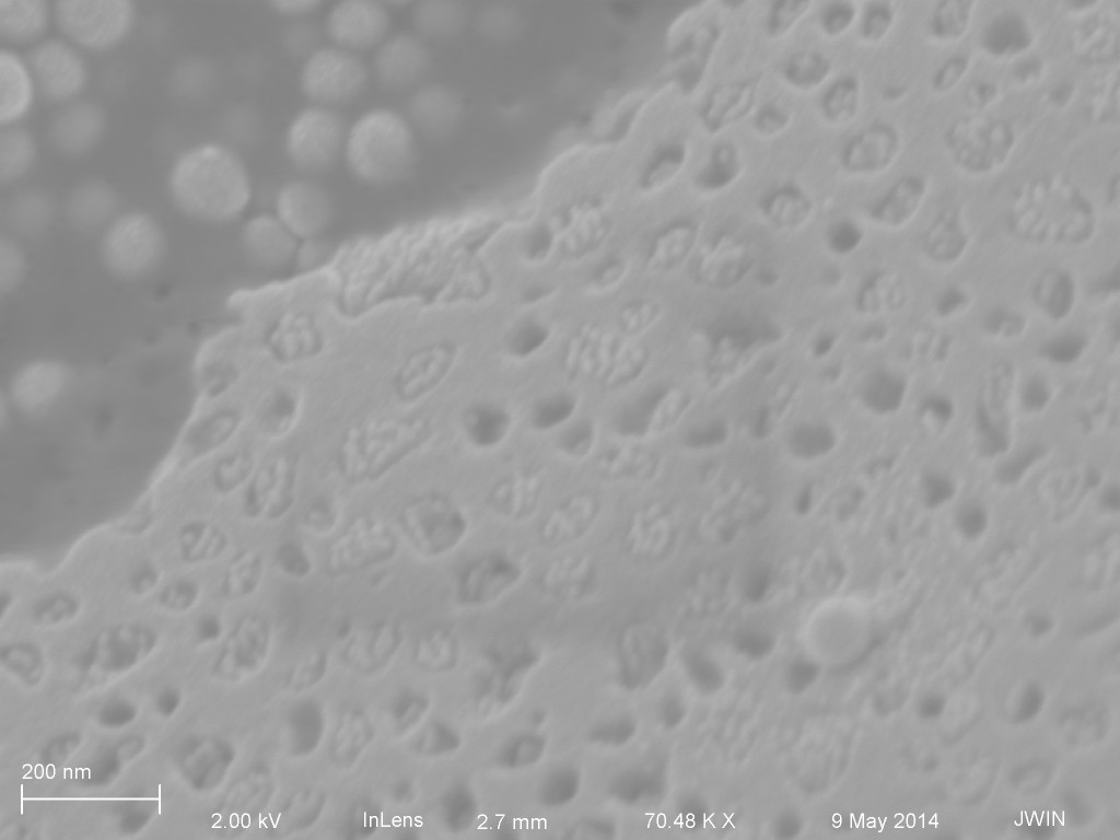
Looking closely at the pores, something that is not 100 nm NPs are filling the pores.
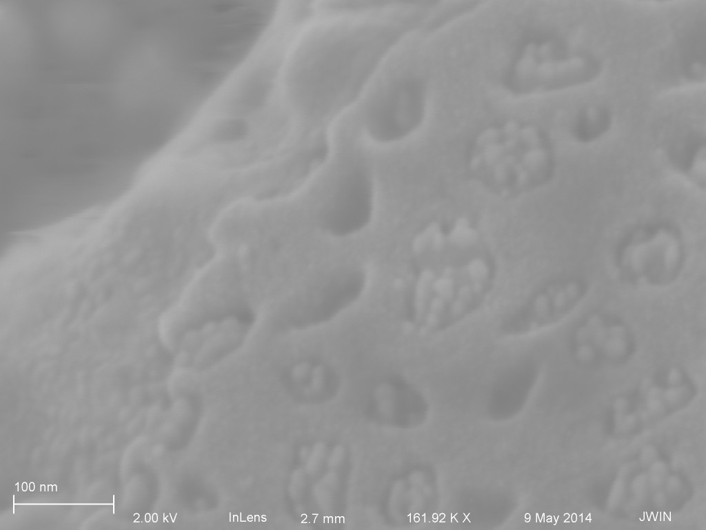
Below are a couple SEMs showing what look to be filled pores. From what I can tell however, this sample was only test with water or PBS. It may have had surfactant as well.