ADSC Proliferation with Different Medias
Overview:
This experiment was conducted to compare the proliferation of adipose derived stem cells (ADSCs) when grown in different medias. Two medias were used and each had different growth factors and serums. Only 0.5um low porosity membranes were used for this study. Data was collected on days 1, 2, 3, and 6, and was used to compare the growth rates of the ADSCs in each of the medias.
Methods:
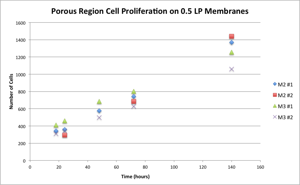
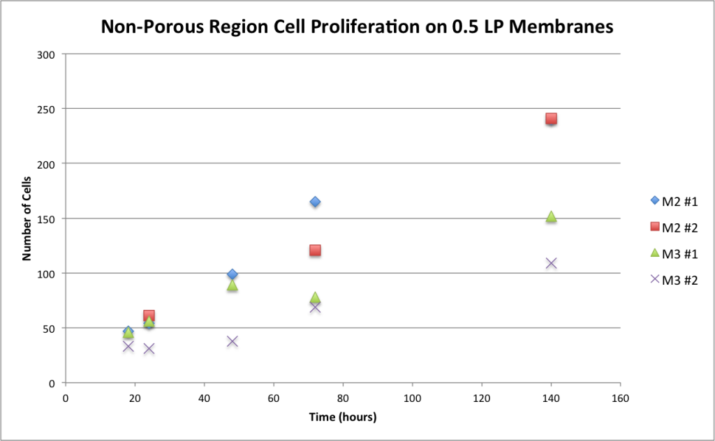
For this experiment, only 0.5um low porosity membranes were used to build CytoVu styled gaskets in a 24-well plate. Each of the membranes was exposed to UV for one hour after being built. A 1% GelTrex in PBS solution was added to each of the membranes for 30 minutes, the solution was then removed and allowed to dry. The cells were passaged and seeded onto each of the membranes at 700 cells/well. The cells were added to each of the membranes in the standard ADSC proliferation media. At the one hour mark, each of the wells were flooded with the media designated for each specific well. Two wells had M2 added and two wells had M3 added (see ingredients below). One of the M2 wells was cultured approximately one month before the remaining three wells. Each of the wells were imaged at 24hr, 48hr, 72hr, and 140hr. Four images were taken of each membrane to completely capture the porous and non-porous regions of the membranes. The cells were counted in each of the images to determine the growth rates of each of the wells. Once all of the data was compiled graphs were made to show the difference in proliferation between each of the medias.
M2 Ingredients- M200 + 2% LVES (standard HUVEC proliferation serum)+ 50ng/mL VEGF+ 1% P/S
M3 Ingredients- M200 + 2% FBS + 1% P/S
Data:
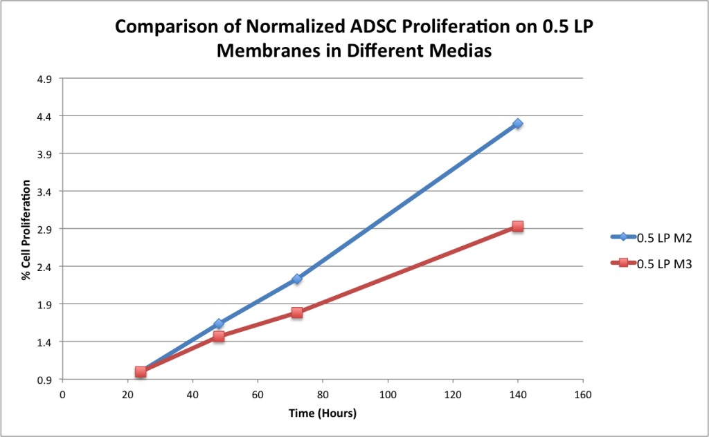
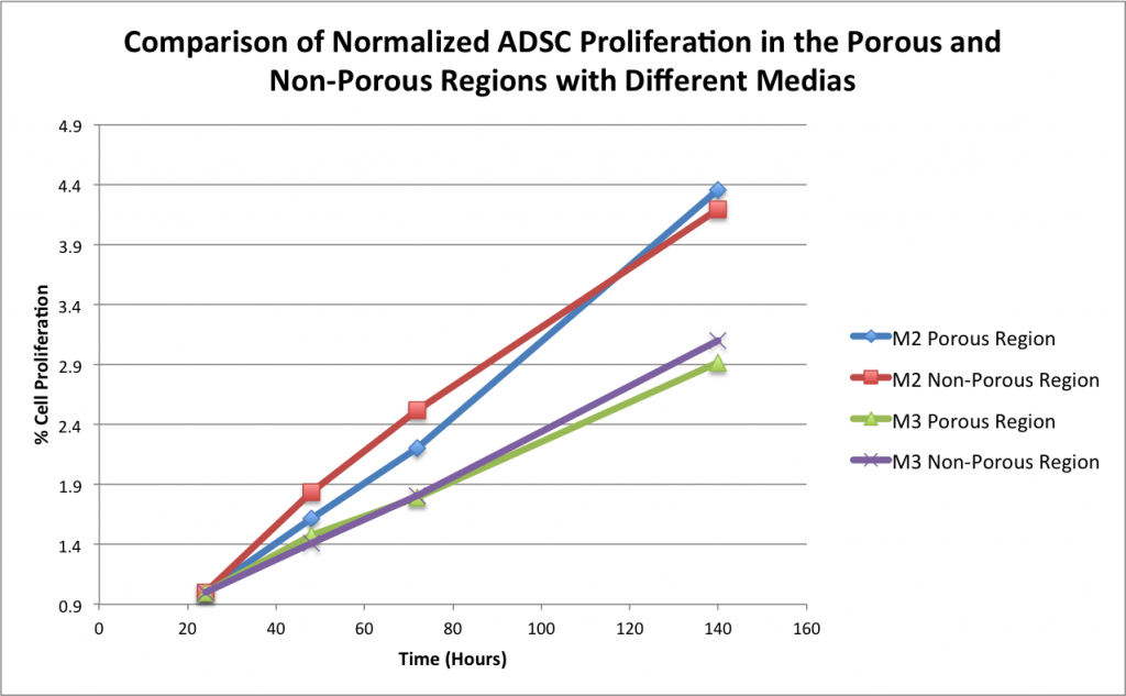
The graphs below were created using the cell count data from each of the two media conditions at 24hr, 72hr, and 144hr.
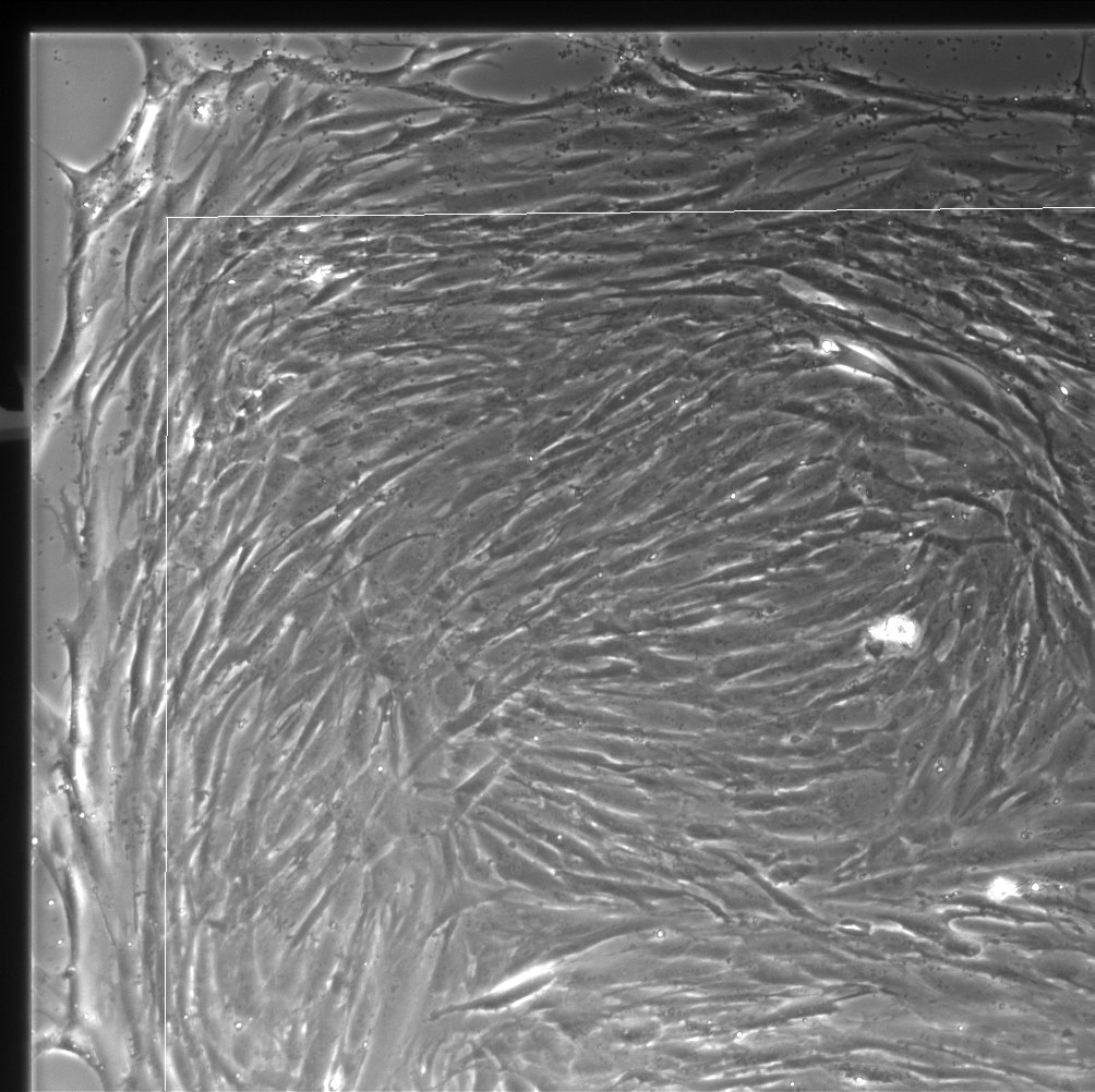
Series of images showing change over time (white line indicates porous area):
24hr, 10x

48hr, 10x

72hr, 10x

140hr, 10x
Conclusion:
The data shows that there is a correlation to cell proliferation and media type. Both of the membranes using the M2 media had greater proliferation by day 6 on both the porous and non-porous regions of the membrane. With the M2 #2 well being from a different set of experiments,the data has even greater support as it proves to be repeatable.