Reversible nano-confinement with a post-filter
My last experiment did not go as planned, but the results are very interesting nevertheless, and might provide another interesting application for these tw0-membrane systems that we had not yet considered.
The start of the experiment went as planned: painted PDMS on the chip, used additional PDMS to glue the gasket to the PDMS layer, which seemed to fix the gasket leaking problem, wetted it, and made a pore. Prior to fabrication, it looked like this:
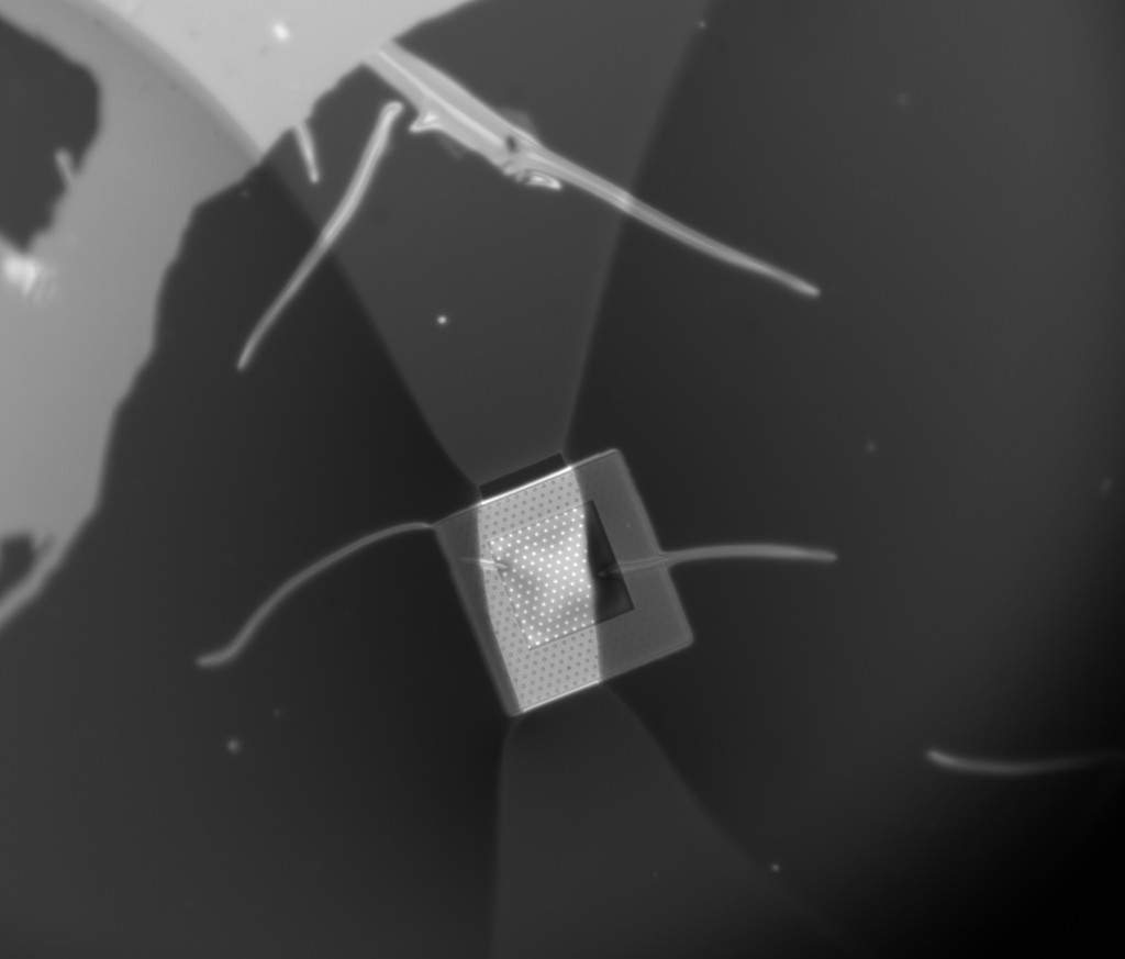
There are a couple of wrinkles, but they are covered by PDMS so I don’t think they matter. For some reason I was unable to wet the membrane using the freezer and instead had to use my breath.
These particular chips take about 30-60 seconds to fabricate a pore at -18V (referenced from the etch bit side of the membrane). One odd thing about this chip was that it had a capacitance in 1M KCl of 1.5nF, which is typical of unpainted chips, compared to the usual ~30pF I would expect for a PDMS painted chip. I have no good explanation of this yet. It’s possible that the liquid was able to get underneath the PDMS layer and contact a wider area than was exposed by the PDMS. I speculate that the extra layer of PDMS used to stick the gasket to the chip caused some stress that partially lifted off the PDMS and allowed liquid to get underneath. This would also explain why wetting in the freezer didn’t work, since I have observed that when a non-sealed gasket is present, vapor prefers to wick under the gasket over staying on the membrane to wet it.
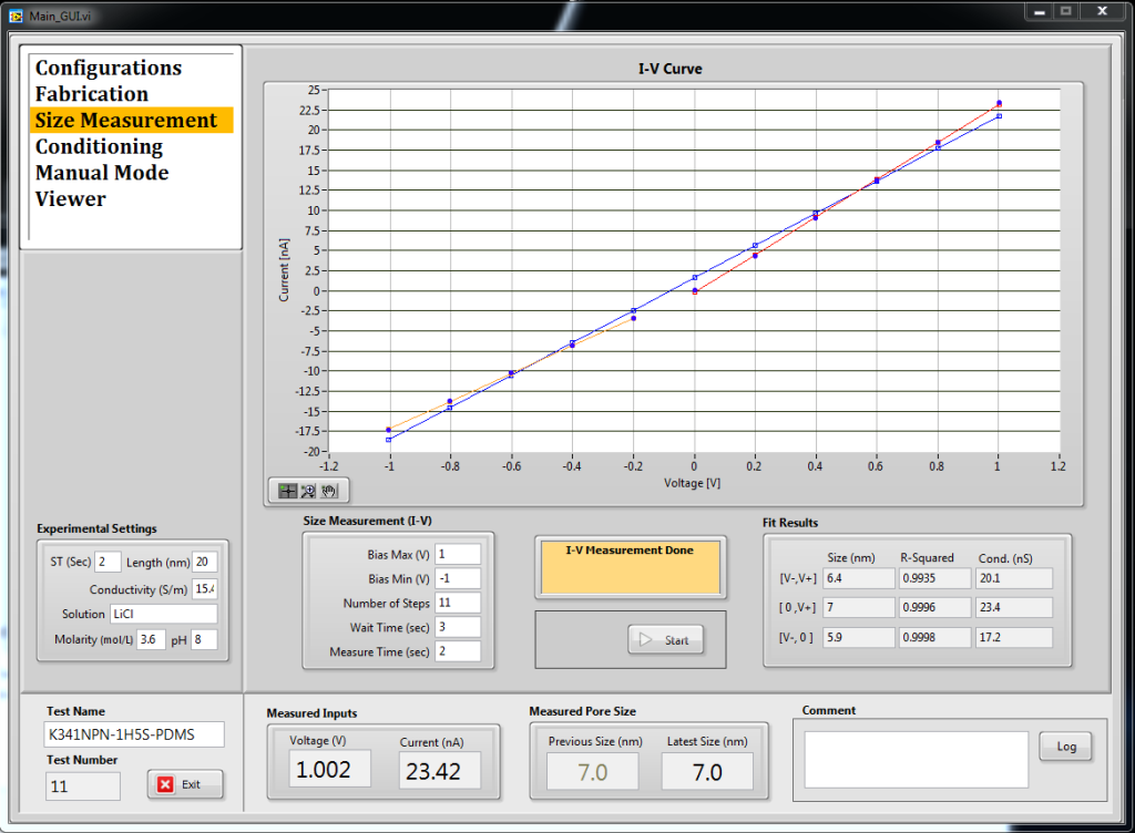
This particular pore stubbornly rectified and had to be conditioned pretty heavily to get it into usable shape, but the eventual IV curve looked like this:
The noise was very good in the low frequency range, which is almost always a good sign for DNA experiments, but because of the weirdly high capacitance it still had lots of high-frequency noise and the peak to peak was less than spectacular.
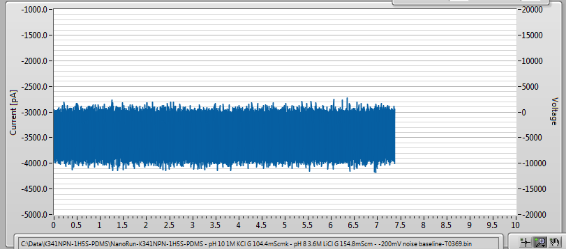
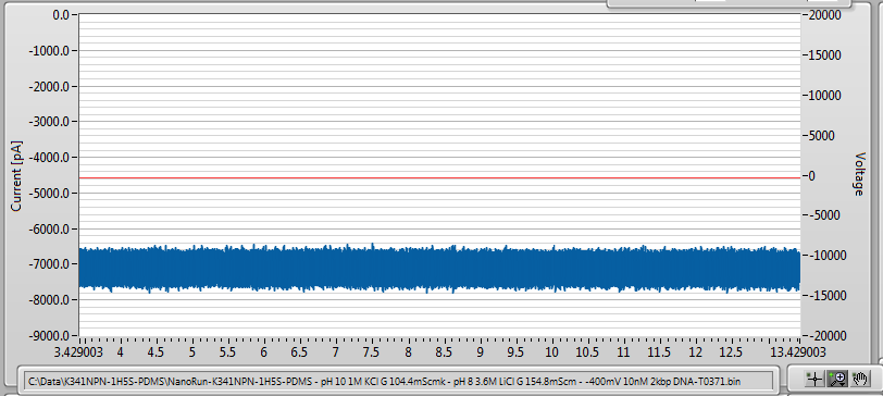
I added 10nM 2kbp DNA to the filter (cis) side and applied -200mV and -400mV. I saw only a single event in several seconds:
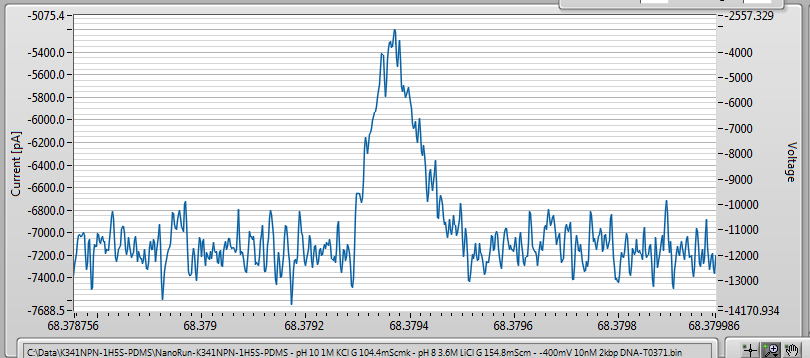
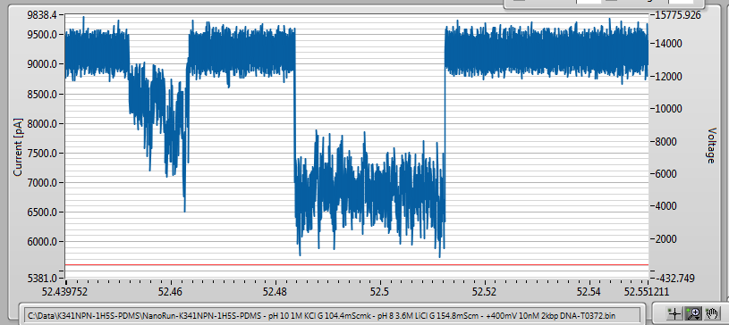
In order to determine whether the pore was defective somehow, or whether it was the filter itself that was preventing translocations, I added DNA to the trans side, with the idea of translocating DNA from trans-cis to see if the pore could pass DNA. Sure enough, I saw events, which means that the filter was the reason for the lack of events. This is unfortunate. However, the events I did see were hundreds of milliseconds long, compared to hundreds of microseconds I would expect to see for this length of DNA in the bare pore case. Clearly the filter is affecting kinetics.
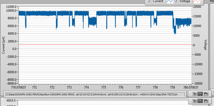
Over time, the event rate got more and more frequent, until events were overlapping and clogging set in. I reversed the polarity to try to clear the clog, and I saw this:
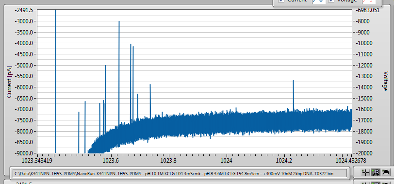
10 or so events, going in the cis-trans direction, which I did not see before. So I tried to reproduce it. Sure enough, while the pore was clogged in the trans-cos (positive polarity) direction, switching between the two polarities would regularly capture 2-10 vents going cis-trans as soon as the voltage was made negative:
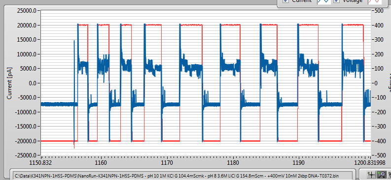
Zooming in on these events, we see that they resenble events I saw earlier where we speculated that proximity to the edge of a well would lead to odd translocation kinetics:
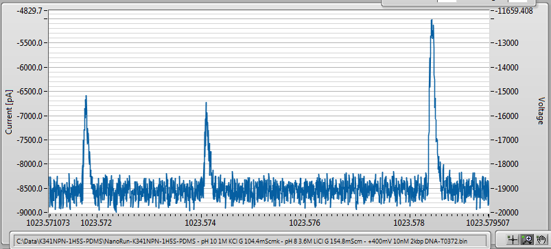
They don’t really look like typical DNA events since they don’t reach a clear plateau, but rather follow an asymmetric pulse shape. This needs further explanation.
Here is what I think is happening:
For some reason (possibly because I wetted the membrane with breath, possibly related to the position of the pore relative to the microwell, possibly because the NPN does not laminate well to the oxide spacer and is too far away from the single pore to experience any electric field pulling DNA through it…) DNA cannot get through the filter, but tends to clog it instead. WHen first translocating from trans-cis I see very long events, indicative that there is some barrier to translocation, which I attribute to the entropic barrier of stuffing DNA molecules into the 200nm gap between the two membranes. As the gap fills up, eventually the single pore clogs since the entropic repulsion balances the voltage-driven addition of new DNA to the gap. Now, if I reverse the polarity, I pull trapped DNA back through the pore. Not all of it, since the pore remains clogged quickly after loading, but the most recent few molecules get reversed and pulled back through.
I have about 30GB of data wherein I simply switch the polarity of the voltage ever few seconds (manually, because involving the computer control adds too much noise to the system). I have not yet analyzed it yet because It’s going to be a pain to do so, largely due to the fact that most of the events happen during the capacitive decay of the current due to voltage switching. The odd pulse shape we see for cis-trans translocation also requires a more quantitative explanation, since proximity to the pore is a little unsatisfying. Usually, I see 2-10 events going from cis-trans before they stop happening and I need to reload the intermembrane gap.
Here is an image of the membrane after doing all of this:
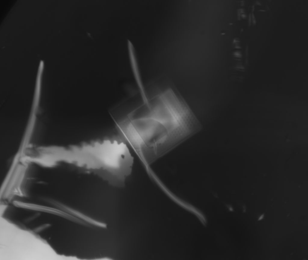
I apologize for the poor image quality – imaging through a liquid droplet is not easy in our system. But it is clear that the NPN layer is still there, which is really all I wanted to demonstrate.
This has some potential uses we hadn’t considered before. Reversible nanoconfinement is an interesting idea that has been approached very differently by other groups. On of the big challenges to electrically confining molecules is the need for an electrode in the nanocavity, but because the upper membrane in our case is conductive, we have no need for that. We only need to sure that the pores in the NPN are small enough to ensure that molecules cannot pass, while ionic current can. In this way, we allow nanoconfinement using remote electrodes and a fairly simple fabrication approach. We could even use this as a reverse filter, if for some reason you wanted to look only at large molecules in a sample: have a single pore large enough to pass your analyte and an NPN layer with pore small enough to prevent it from escaping the inter-membrane gap while passing any small contaminants. Then the first few events observed upon switching the polarity could reasonably be assumed to be due to the larger molecules only.
After doing this experiment at 200, 400, and 600mV I flushed it out and try to spike in 20kbp DNA to dot he same thing. I was able to see events, but at fist glance they seem not too different from the 2kbp ones, suggesting that the inter-membrane gap is still loaded with hundreds of 2kbp molecules.
Assuming a radus of gyration of about 75nm for 2kbp DNA, the gap will fit around 350 molecules before filling up, call it 100-1000 since the model is very crude.
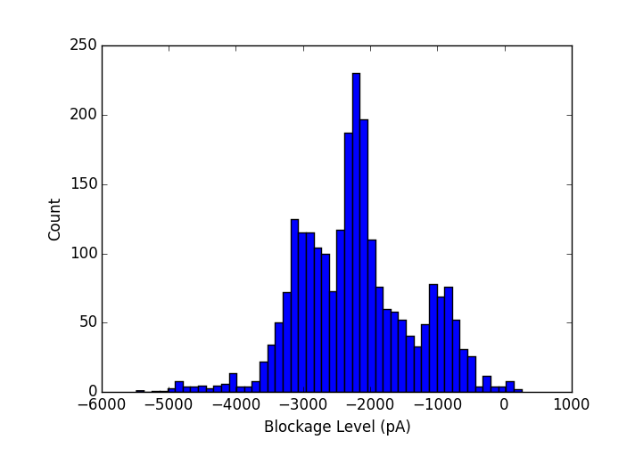
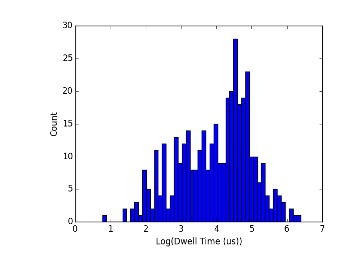
Counting the number of molecules in the gap, the first permanent clog I experienced (there were a few reversible ones prior) was after about 900s of data. In that time, I collected approximately 656 events of which my analysis code accepts 383 as being real translocation events, so either way we are in the right ballpark for for closely packing the entire oxide spacer gap with DNA molecules.
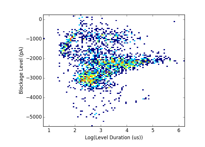
Some statistics show extremely long dwell times (note log scales). Blockage depth expected for dsDNA in these conditions is about 1180pA, so the main peak appears to be doubled DNA translocations either folded or coupled. Dwell times run into the seconds
So, not really what I was going for, but sometimes accidental experiments are the best ones. Let me know what you think. I still have some work to do to analyze the captured translocations from cis-trans, but that might take me a little while.
EDIT: Vincent pointed out this paper, which uses nano-confinement but with a two-pore approach:
Very interesting! Thanks for writing such thorough posts. Its the only way I can follow.
I mistakenly thought we were working with smaller DNA molecules than this. Is there really any surprise that the cis-trans experiments are slowed down if the DNA has a radius of gyration of 75 nm and the NPN pores are 35 nm? The membrane isn’t necessarily clogged (for that I need to know the DNA concentration – so what is it?) but certainly individual pores are blocked. The DNA must unwind as if at the end of a spool of yarn to reach and enter the pore some 200 nm away. Such events should be slow and infrequent, right? By the way, this is the picture Vincent drew for us back in the early days of this project. I’m not sure if he anticipated slower kinetics, but it makes sense (to my half-informed brain anyway). If it is worth working with smaller DNA that fly through pores, I don’t know. But presumably the pre-filter would do its job there. Perhaps this is changing the publication target for us?
Nanoconfinement is very interesting and a wonderful offshoot of this work. I uploaded the paper Vincent referenced. Are there clear applications for nanoconfinement? Or are people currently just studying the cool physics?
Yes, I would expect slower kinetics for sure, which is why I was surprised in my last success in which it appeared that DNA was able to get through more or less freely. I suspect that it has a lot to do with exactly how far the NPN is from the hole. At 200nm, there is probably a small but significant pulling field across the NPN (maybe a few mV) whereas if it balloons out much we would be relying on pure diffusion.
I think it’s worth looking at a range of sizes. 100bp, 1kbp and 10kbp or something like that, to see differences. Problem is that multiple samples in the same pore, sequentially or otherwise, is very difficult. But I’ll keep trying.
The DNA concentration I used is (always unless the experiment specifically requires something else) between 1nM and 10nM.
Nanoconfinement has potential uses as a way to reduce speed variations along the length of a molecule by limiting the number of initial conformations that DNA can adopt before translocation. That being said, 200nm x 1um confinement is really not very much, so for this form factor I would not expect to see much in that regard. But being able to capture, keep the DNA localized on the other side, and pulling it back through is probably worth exploring.
Sorry. Concentration isn’t what I need. How many molecules of DNA are you putting in the system? Assuming they all get pulled to the NPN, I would like to compare to the number of pores.
I’m not sure I understand – there are 35 microliters of solution at 10nM means ~2e11 DNA molecules in the system. If you mean how many actually got into the inter-membrane gap, the answer is that I detected about 383 events that I am reasobly sure are translocation events before it permanently clogged, but it should be noted the the clogged state is only about 45% lower in conductance than the open state, so it’s not a complete clog. It’s also likely that the clog is happening at the single pore, since that is the highest resistance in the system and therefore more likely to register molecules in its vicinity than the NPN.