YAP/TAZ Project Summary
First off, this is the protocol for the staining and analyzing the data is shown below and was used for all of the YAP experiments:
For the substrates, we wanted to use 0.5μm HP, 0.5μm LP, 3μm HP, TCP, and nonporous SiO2. Cyto-Vu devices were made using the aforementioned substrates and 300 ADSCs were seeded per membrane. The cells were allowed to adhere to the substrate for 1 hour and then the wells were flooded with 800uL of media. After 3 days, the cells were fixed in 3.7% formaldehyde for 15 minutes, permeabilized with 0.1% Triton T-100 for 3 minutes, washed with 4% BSA for 15 minutes, and 100μL of the YAP antibody (0.2mg/mL was diluted 1:100 in PBS) was added on top of the substrate for one hour.
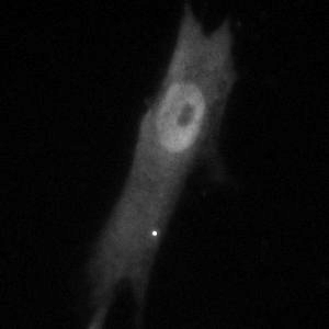
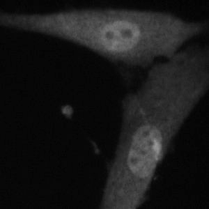
The images were acquired and analyzed in image j. The nucleus’ brightness was measured as well as the background and the cytoplasm of the cell. To analyze the data, the background was subtracted from the values for the nucleus and the cytoplasm. Then, the intensity of the nucleus was divided by the intensity of the cytoplasm. Sample sizes were at least 20 cells.
To first look into our YAP data, we wanted to look at the spread area of our ADSCs just to make sure spread area wouldn’t be impacting the YAP expression because literature shows that it can. To do this, I took the YAP images from the 24 hour passage 1 experiment and the threshold was adjusted on the images until the cells were entirely white and the background entirely black. From there, I used the “magic selection” tool to capture the size of each cell. A Tukey test was done in Minitab to analyze the results and cells over 6000μm and under 3500μm were considered outlying and were eliminated.
After an analysis of the samples (after removing outlying values) there are no statistical differences between any of the substrates with a 95% confidence interval. This data is similar to the ADSC spreading data that I collected two summers ago, the means/error bounds are relatively similar.
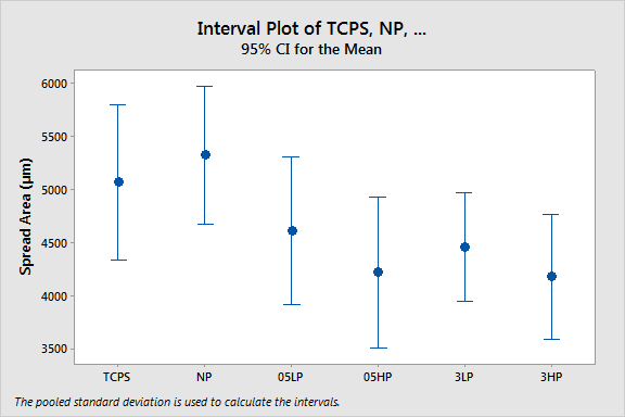
Confirming that our spreading data was different, slightly, in an insignificant fashion, we can rule out any major impacts of spreading on YAP expression.
From there, we went with literature and stained for YAP after 24 and 72 hours (we did not see much difference between the two time points). To rule out possible substrate memory influences, we first did our experiments with passage 1 ADSCs (On TCPS for two days) and repeated the experiments after 10 days to see if we observed any sort of mechanical memory from the ADSCs.
Here is the data for our P1 experiments after 24 hours and 72 hours respectively.
24 Hour:


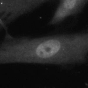
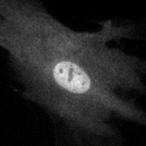
Representative Images (40x)
3μm HP 3μm LP 0.5μm HP

0.5μm LP Nonporous SiO2 TCPS

72 Hour:

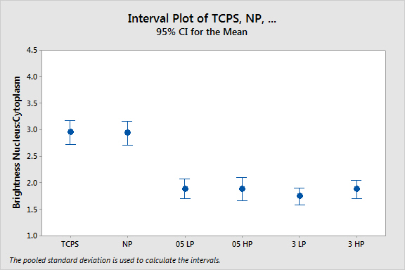
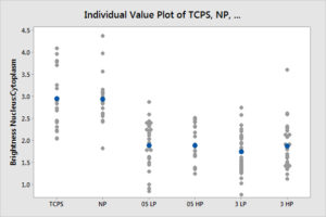
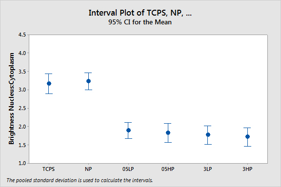
From 24 to 72 hours, we see no difference in trend and minimal differences in the means of each substrate. Data from both days shows that YAP expression between TCPS and Nonporous SiO2 are not significantly different and between our porous substrates, it isn’t either. On the converse, the porous substrates are significantly different from the nonporous substrates. This would indicate that the membranes are being perceived by cells as softer substrates, possibly due to the broken up surface area. This could indicate possible pathways for YAP expression and that continuous surface geometry can impact how cells perceive a substrate as soft or stiff. It’s interesting to note that there aren’t significant differences between any of the porous substrates despite varying porosity or pore diameter.
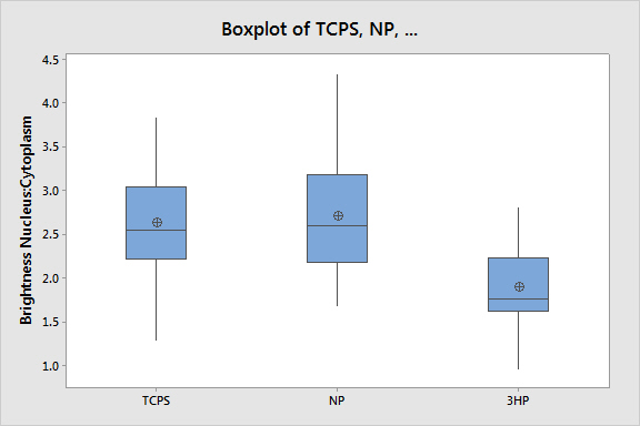
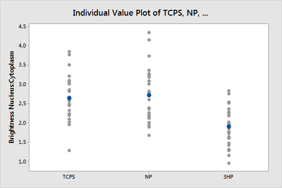
Just to investigate memory further, we tested cells on our most polarizing substrates (TCPS, nonporous glass, and 3μm HP membranes) to see if the YAP localization was impacted in comparison to the passage one ADSC results. These ADSCs had been “treated” (dosed) with TCPS for 10 days, which in literature would inhibit cytoplasmic YAP localization. The results are shown below:
24 Hour:

72 Hour:

These experiments would show that contrary to literature, we aren’t observing the same memory affect that some other groups have seen. This is good for our experiments because it shows that passage one cells are not necessary to collect particular sets of data.
Summary:
- Spread area for ADSCs is not significantly different between porous and nonporous substrates
- YAP localization is similar on porous substrates despite varying pore size and porosity
- YAP localization on nonporous substrates are similar
- YAP localization is different between porous substrates and nonporous substrates
- ADSC “memory” was not observed