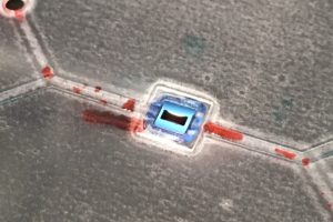
Fisher @ NRG, Summer 2017
We are happy to be back this summer. Anthony has been working on setting up a system that mimics Kilean’s dialysis devices.
Our approach is different in two important ways.
First, we are using PETLs, instead of PDMS/glass. For those who are not familiar with PETLs, here is a short summary:
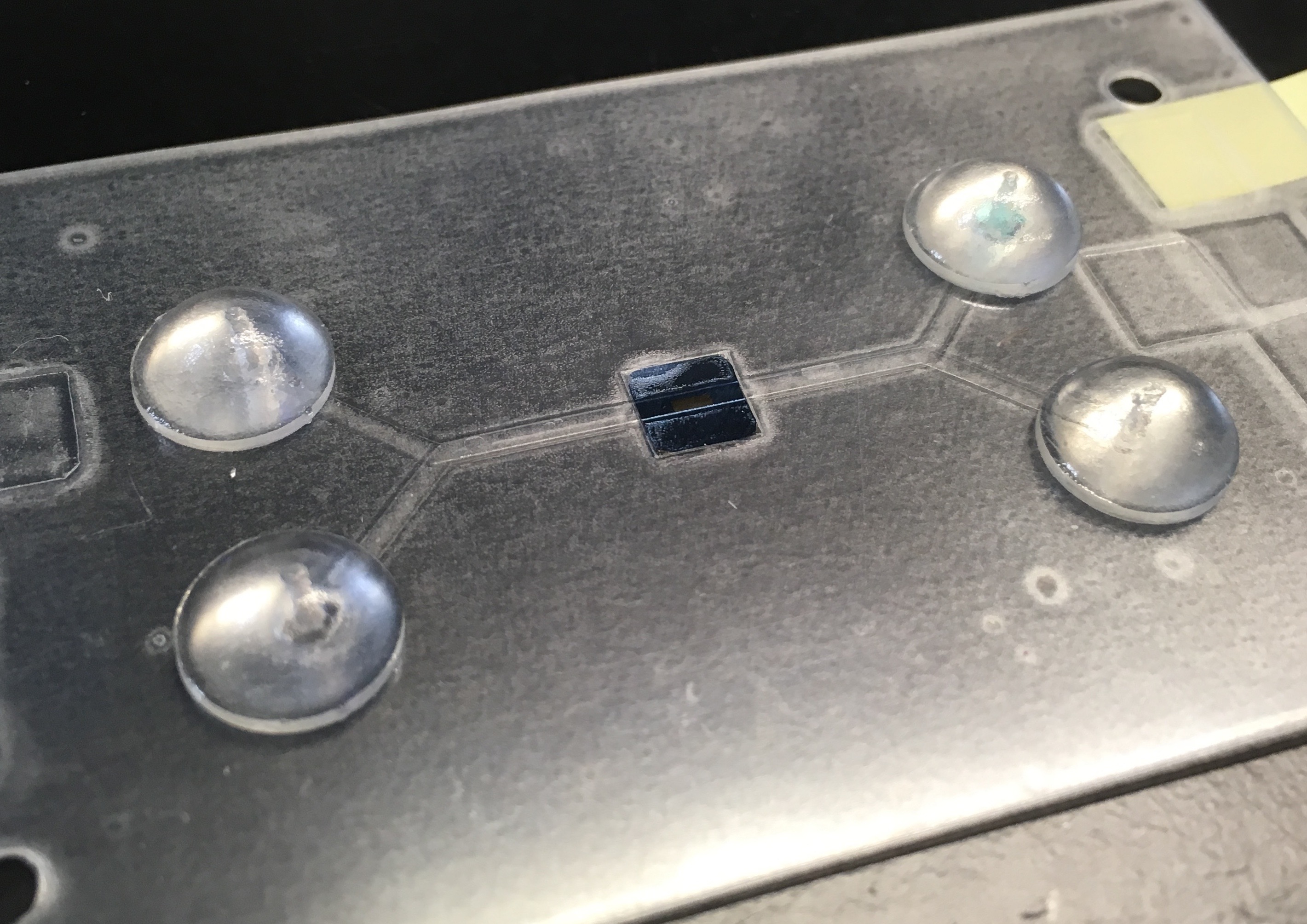
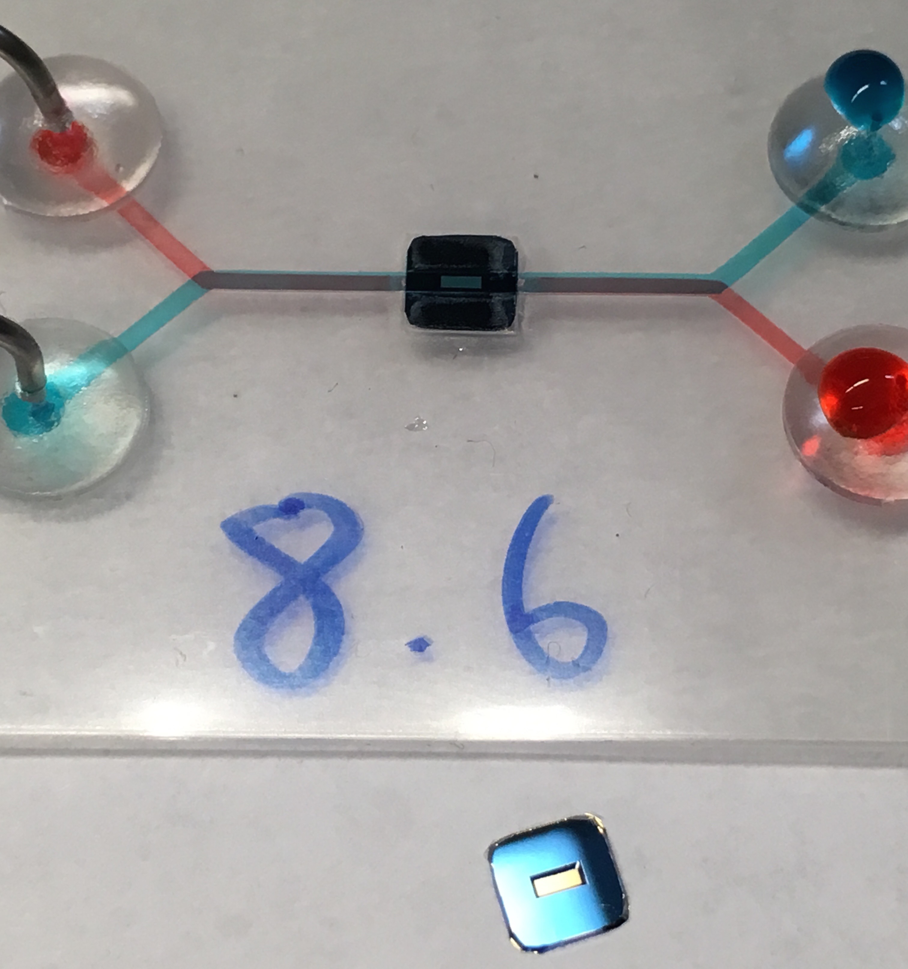
Commercial laminating pouches made of PET film are coated with a thermoadhesive called EVA. We use a craft cutter to cut the channels in single film layers and then sandwich and laminate them to seal the device. The thickness of the film determines the height of the channels (usually 3mil=76microns). The width of the channel is determined by the user, with a ~150-200 micron lower limit imposed by the craft cutter. Acces to the channels is facilitated by self-adhesive, perforated vinyl bumpers.
For more info and a video on PETLs go to https://sites.google.com/a/sjfc.edu/ontiveros-lab/
Second, our goal is to detect viruses, instead of exosomes. Most viruses go from ~40nm to a couple of hundred microns in size.
Our aim is to catch viruses that approximate the size of the pores, much like Kilean does with gold particles and exosomes.
So the project is straight forward. For the last 4 weeks Anthony has been working on the chip’s architecture and also training on the SEM.
We think we have a winner design, and will be imaging it (after testing with gold particles) tomorrow (6/29).
If things look good with gold, we will start designing experiments with viruses. Stay tuned, it will be a fun 6 weeks ahead.
Some comments on the PETL design we will use:
a) The channels are 76 microns in height, and about 1100 microns in width. The width was designed to match the membrane’s window on both sides.
b) The device is made of 9 superimposed layers. The total thickness is under 700 microns.
c) The layers are cut in the silhouette and bonded using a laminator (~130C); the cost is under a dollar per device (not counting chip) and fabrication takes under 1/2 hour.
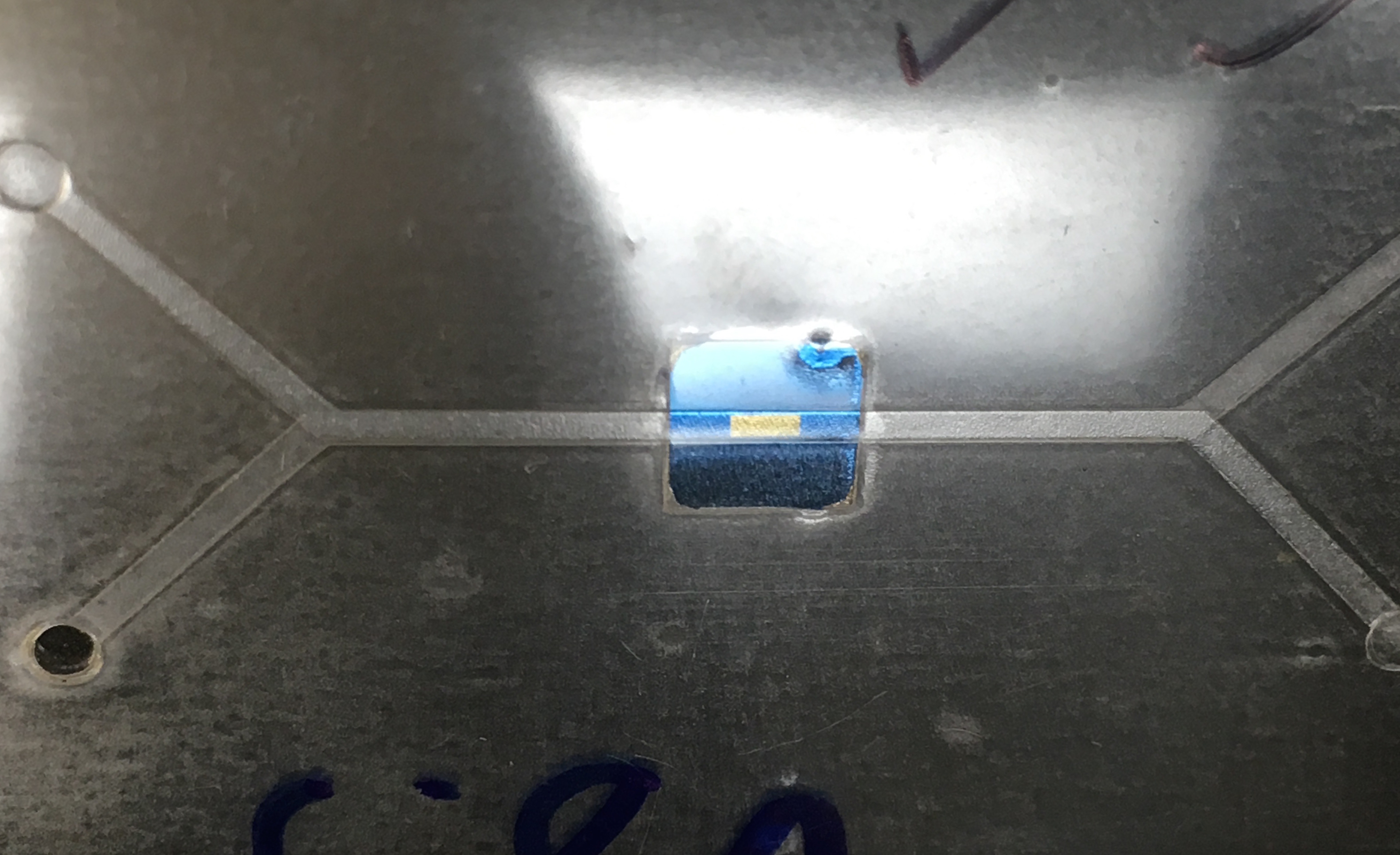
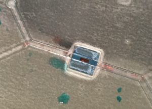
d) The top and bottom layers can be separated (delaminated) after use (see pictures), to facilitate access for imaging.