Exosome Scale Experiment (Summary)
Hello Everyone,
So following up on my exosome scale experiment and my last post, i am presenting my last update to summarize my results and to show that my results are trustable and reproducible.
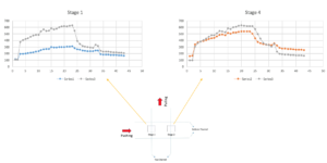
This is how we take images:
We have different positions in the interaction area, and we take images as the experiment is running. Different regions of interest from each image will be picked and the fluorescent intensity will be measured. Then the results for different regions of interest and images will be analyzed.
This is how we analyze our data:

Same experiment was repeated couple of times with successful repeatable results. In all of the experiments, the sample was pumped by 5 microliter/min in bottom channel and pulled from the top channel by 2 microliter/min, and captured beads were released by 4 microliter/min from the tip channel through the membrane to the bottom channel.


Results from another experiment:
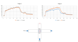
In this experiment, we decided to check the intensity of 2 stages that are away from the interaction areaas you can see in the figure.

The next step in my experiment will be checking consistency of my results, but in order to check that, i need to make sure that my experiment condition is the same in all experiments like volume of sample, experiment time, concentration of beads in the sample. If we see the consistency in the results, we can move forward to change the experiment condition to see the effect of different parameters such as albumin and serum to decrease the non-specific absorption. I would like to check by SEM and make a rough estimate of number of captured and non-specific absorption and compared this result by our analyzed results. Also, I have been working on a new set of experiments with mixture of beads with different sizes to show the possibility of capturing particles of interest and releasing them while smaller and larger particles are getting filtered or washed away.
So I wanted to see if the green fluorescent beads would contribute or leak to the red signal, so ran one of my experiments with 100nm green beads over 80 nm pore size TE membrane and i imaged with both GFP and TX-Red filters. The good news is that we don’t see any effect of green beads on Red signal, and the figure shows successful capturing and releasing on GFP which was expected, but a plateau for TX-Red. I am planning to check the other way around and a mixture of samples with 50 nm red and 100 nm green beads.
