As a continuation to my previous post, I am running a triplicate of cell culture adhesion tests to observe cell lose on our membranes following physiological shear stress.
Methods
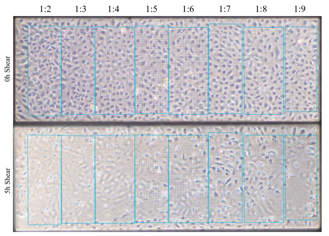
HUVECs (P3,4,5) were seed within our mimetic and left to grow to confluent for 24 hours. Shear (4.5 dyn/cm2) was initiated and images were collected following 5 hours.
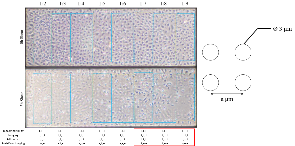
Figure 1. Preliminary data on cell adherence to μ-NPN material. Cell loss in the first 5 hours is most prominent in the first three regions (1:2, 1:3, 1:4).Figure 2. Data collection and characterization. Results were binary and collective result was used to base my final membrane group selection (n=3).
Figure 3. Heterogeneity in shear results a function of pressure?
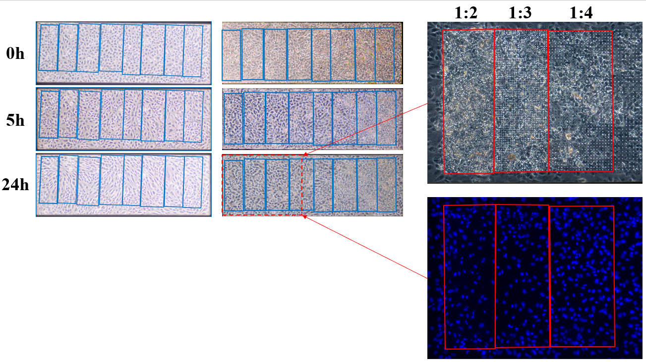
Figure 4. Cell loss under cell material in side by side pattern experiments.
A quick update since we last talked about how DNA controls were proving to be problematic: Things are working again (more or less). I went back top basics, remade most of my solution, ordered new DNA samples, and, most importantly, piranha-cleaned my chips. The first three experiments I did on basic 30-nm thick 5.4mm square…
We’ve completed two successful 4-hour small animal hemodialysis experiments. I’ve also analysed the serum from all previous experiments. This graph shows only the recently analysed serum and doesn’t include the previously analyzed data. I’ll give a more detailed analysis of these results when we finish the next two animals, mid April. I’m in the midst of submitting…
I spent some more time in understanding the numbers I am getting for our device simulations. In my simulations, I am assigning a conductivity value for my cell layer. Technically, from this information and by knowing the thickness and area of the cell layer, I can analytically calculate the value of resistance that the cell…
A couple weeks ago when I presented at lab meeting, Angela Glading suggested that I look at epithelial cell vacuolization on transwells. This was in order to determine if vacuole formation was endothelial cell-specific. If I’m remembering correctly, the rationale was that the vacuole-less fibroblasts I showed were not contact-inhibited cells and thus not a…
Thanks to SiMPore’s push for TEM grids, we’ve produced a lot of amorphous material in the last month under different deposition conditions. Below I’ve illustrated the different film structures resulting from depositing with and without substrate bias and heating. Without bias, the amorphous film exhibits clusters of thicker/denser? material (darker spots). Intuitively, this makes sense…