Polarized TNF-α Update: rhIL-8 and PMN Persistence
Introduction
In a previous post I introduced the concept of endothelium apicobasal polarity and the potential to utilize this concept for future sepsis drug development: Endothelium Apicobasal Polarity Regulates the Innate Immune Response In Vitro [In Progress]
In this post, I will expand on the ‘mock’ TNF-α experiments with recombinant IL-8 as well as add details on some of the observed neutrophil (PMN) dynamics.
Results and Discussion
Recombinant Human Interleukin 8 (rhIL-8) Reverses the Observed Transmigration Response on Stimulated Endothelium
Abluminally stimulated endothelial cells [20 ng/ml TNF-α, 24 h] and control cells were rinsed of spent media and perfused with rhIL-8 supplemented media at concentrations as observed in our ELISA experiments (see previous post for details). PMNs were added into the devices and phase time lapse movies were collected. In the case of the non-stimulated endothelium, no PMNs were observed to transmigrate over the course of the 30 min experiment (Figure 1A). Positive control experiments (2 nM rhIL-8 supplemented abluminally) demonstrated the chemoattractant ability of IL-8. In abluminally stimulated devices, we previously observed a potent PMN transmigration response (Figure 2). Interestingly, while the addition of rhIL-8 at concentrations observed in abluminally treated devices preserved the transmigration response, the same experiment at IL-8 concentrations observed in luminally treated devices completely eliminated PMN transmigration (Figure 1B). These results support the conclusion that IL-8 is partially responsible for the observed transmigration response, and that cases of PMN sequestration in sepsis may be due to a reverse gradient holding PMNs in the lumen.
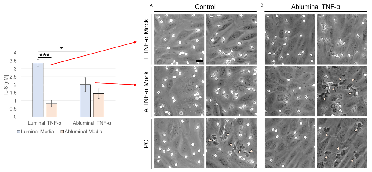
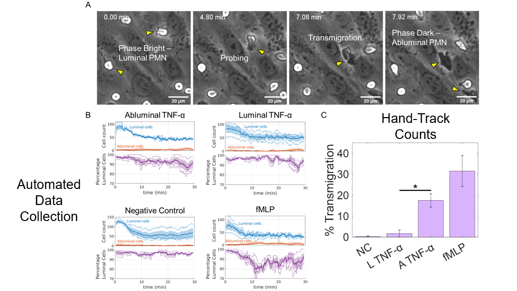
The results presented here are supported in part by previous seminal works. Kim and Haynes (Anal Chem 2012) previously demonstrated the potent chemoattractant potential of IL-8 to neutrophils. Additionally, they concluded that IL-8 is strictly a chemoattractant, and not a chemokinetic, meaning that while IL-8 does drive directional transmigration, the presence of a gradient is necessary. This is supported by our result on unstimulated endothelial cells where reverse and relatively equal concentrations of rhIL-8 did not produce a transmigration response. These properties of IL-8 are presumably why much confusion surrounded the molecule as to whether it is in fact a chemoattractant or an inhibitor of chemotaxis. One result that is unclear under these conclusions is the transmigration response on stimulated endothelium. However, IL-8 had been shown to adhere to the basement membrane in vitro (Huber et al., Science 1991), preserving the chemotactic gradient. This reserved IL-8 may add to the total basal IL-8 concentration in stimulated cultures, thus providing the means of the transmigration response. In the case of ‘mock’ luminal TNF-α experiments under these same conditions, these data suggest an overwhelming amount of IL-8 secreted into the luminal compartment in sufficient to disrupt the gradient and hold PMNs in place. Future work should focus on a mechanism of IL-8 direct secretion, potentially aimed at the ability of endothelial cells to store IL-8 in Weibel-Palade bodies for rapid immune responses. With our platform and the introduction of super resolution microscopy techniques, we believe this work is possible.
PMN Migration Dynamics Correlate With Cytokine Stimulation, Uncoupled from Apicobasal Delivery and Transmigration Response
Neutrophil dynamics have been a research focus since the early 90s, as they represent a group of highly abundant and highly motile innate immune cells that act as first responders in the cases of infection or inflammation. Having a system capable of observing and quantifying neutrophil dynamics on a permeable support, it is clear there is potential to elucidate some interesting neutrophil biophysics in a controlled environment. The results of these experiments are represented in the Figures 3 and 4.
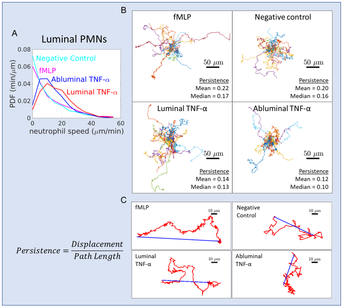
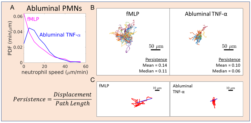
The results show a correlation between cytokine stimulation and neutrophil speed and persistence (ratio of total displacement to path length): When endothelial cells were stimulated with TNF-α, increased speed and decreased persistence was observed compared to controls (Figure 3). This result is expected based on previous studies, but adds to these works by showing how these dynamics are uncoupled from the subsequent transmigration response (as observed in fMLP and abluminal TNF-α groups). These data also further support the claim that PMN transmigration can be mediated by cytokines or chemoattractants, and the mechanisms of both are somewhat divided in nature. Interestingly, when PMN migration dynamics were quantified abluminally (i.e. speed and persistence of PMNs that have already transmigrated), we still observed an increase in PMN speed under stimulated endothelium and a corresponding decrease in persistence (Figure 4). This result suggests TNF-α can modulated abluminal PMN migration as well as the well studies luminal migration. Given we are observing integrin mediated migration in our system (no shear flow = no selectin mediated rolling), this may suggest that abluminal TNF-α may modulated either β2 integrin mediated ICAM-1 migration or β1 integrin mediated basement membrane migration. In searching literature, I found little evidence supporting either hypothesis. Given our ability to label ICAM-1 and image it with confocal, I decided to give this experiment a try (Figures 5 and 6).
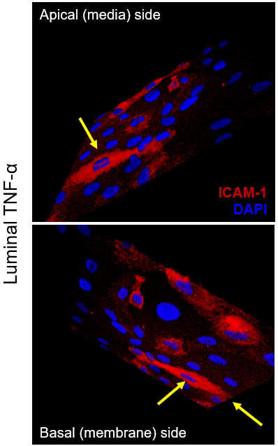
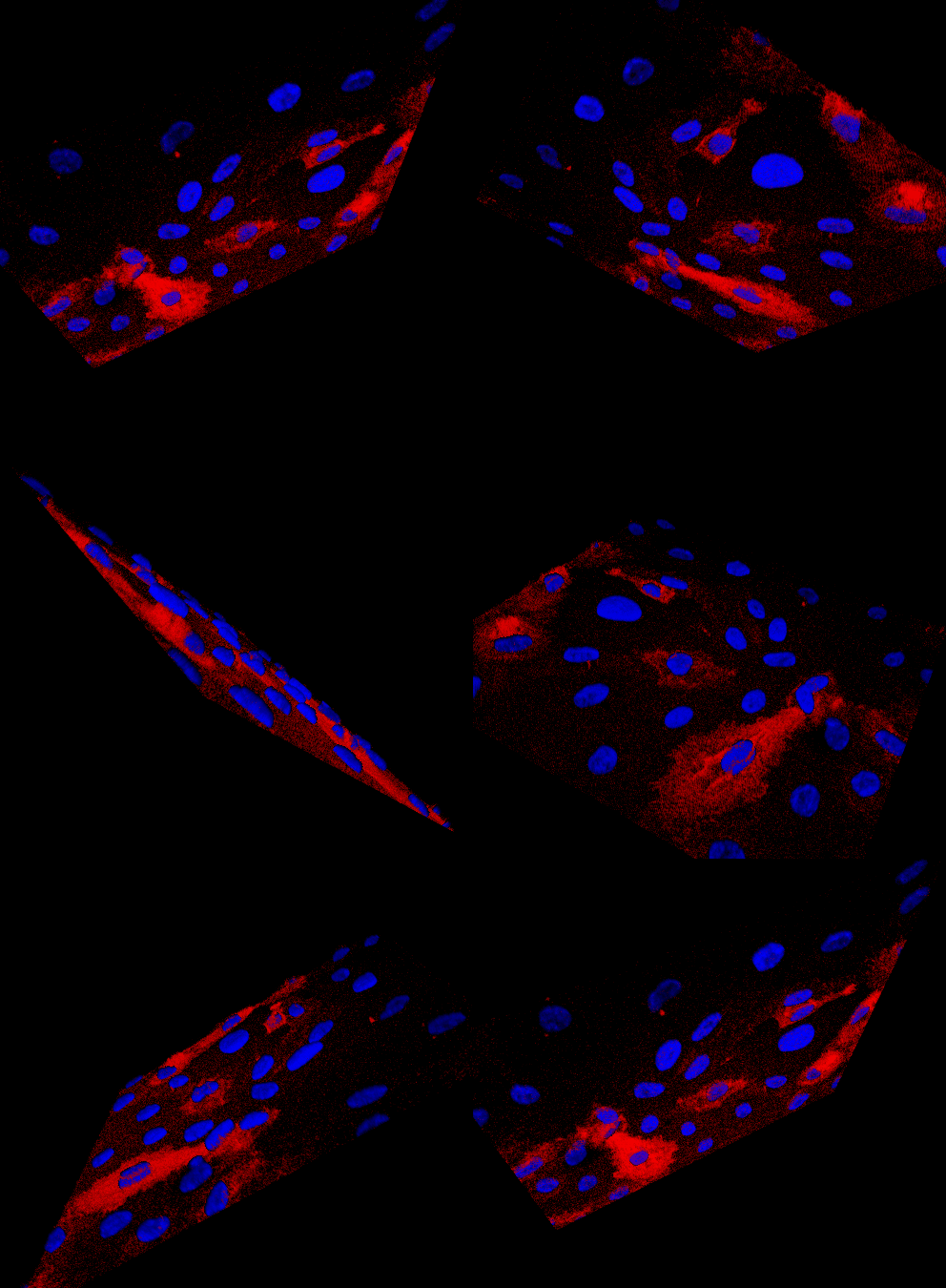
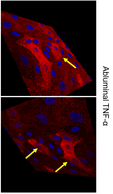
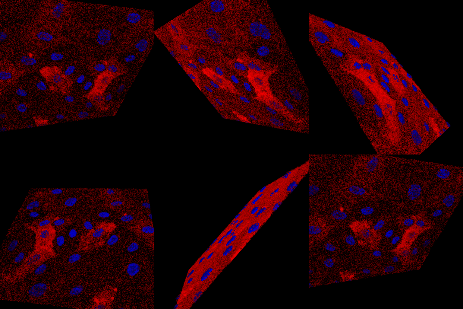
While strictly preliminary in nature, it does appear that there may be increased ICAM-1 stain under the nuclei in abluminal TNF-α treated devices, maybe suggesting increased abluminal ICAM-1 expression. Regardless, it is clear ICAM-1 staining is prominent on the luminal cell surface in both cases. Super resolution microscopy is needed to strengthen these results, but is out of the scope of this work.
Future Directions
More PMN dynamics? More antibody blocking experiments and CRISPR cell modification? Receptor labeling and super resolution? (Will expand later…)