μSlide to hold μSiM devices for enhanced imaging & fluidic applications
Introduction
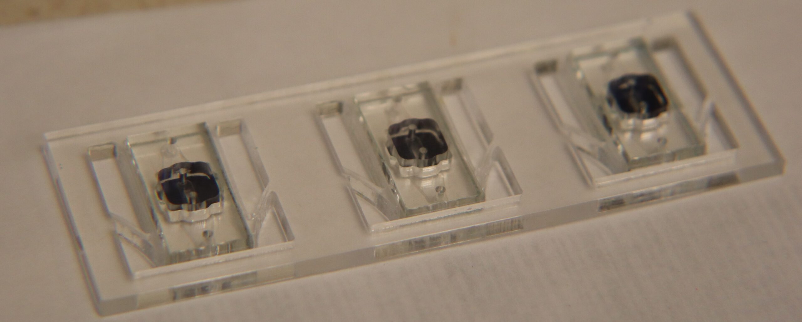
The Slide Body (Figure 1) has been used for a long time to image μSiM devices. This slide body has the same footprint as a traditional microscope slide but is designed to hold up to 2 μSim devices for imaging. The device has an open center with thin ledges where the edges of the μSim device can be held so the area of interest is clear of any obstructions when imaging. However, some limitations apply to the current version of the current slide body. For example, while the ledges that the μSiM devices are on are thin, some working distance is lost making it difficult to use high magnification systems. In an era where we are trying to do super-resolution imaging with structured illumination microscopy having the least amount of materials between the objectives used and the membrane is key to excellent imaging. Furthermore, the ledge where the device sits also poses the risk of not being flat during the manufacturing process of these components. This unevenness of the ledge was quantified by focusing on one corner of a membrane window, recording the z-height of the objective, moving to the opposite membrane window corner, and finally recording the new microscope objective z-height. Across 3 measurements on the same slide body, this showed an average total tilt of 18.1 μm. When the same μSiM device was measure for tilt on a plain microscope slide, the measured change was 1.7 μm. This 1.7 μm tilt comes from a combination of the tolerances within the microscope and the tolerances within he microfluidic device. These measurements indicate that the ledge creates a tilt of 16.4 μm. The same corner to corner tilt test was done with a 3D printed model of the μSlide which showed an average total tilt of 7.8 μm across 3 trials. Accounting for the 1.7 μm tilt of the system, the μSlide showed a tilt of 6.1 μm. The data used and the calculations for these results can be found in Table I below.
*Note (4/26/2024): These calculations are just preliminary and some human error is likely present due to the microscope being used having focusing issues on the corner of the membrane window. Once the focusing issue is fixed, the same experiment will be repeated and the results will be update here.

Figure 1: μSim Slide Body

Figure 1: μSlide CAD Model with uSiM device

Figure 2: Dimensioned Drawing of μSlide.

Table I: Microscope objective z-heights used to calculate tilt.
We have a FormLabs 3B printer in my lab you are welcome to use – we’ve used a lot of resins with it, including the high-temp resin, if you needed to autoclave the slides.
We also have a Gamma Irradiator in the basement of Institute Hall. It hasn’t been used in a few years, but to our knowledge, it should be operational.