Updated PETL Dialysis Images
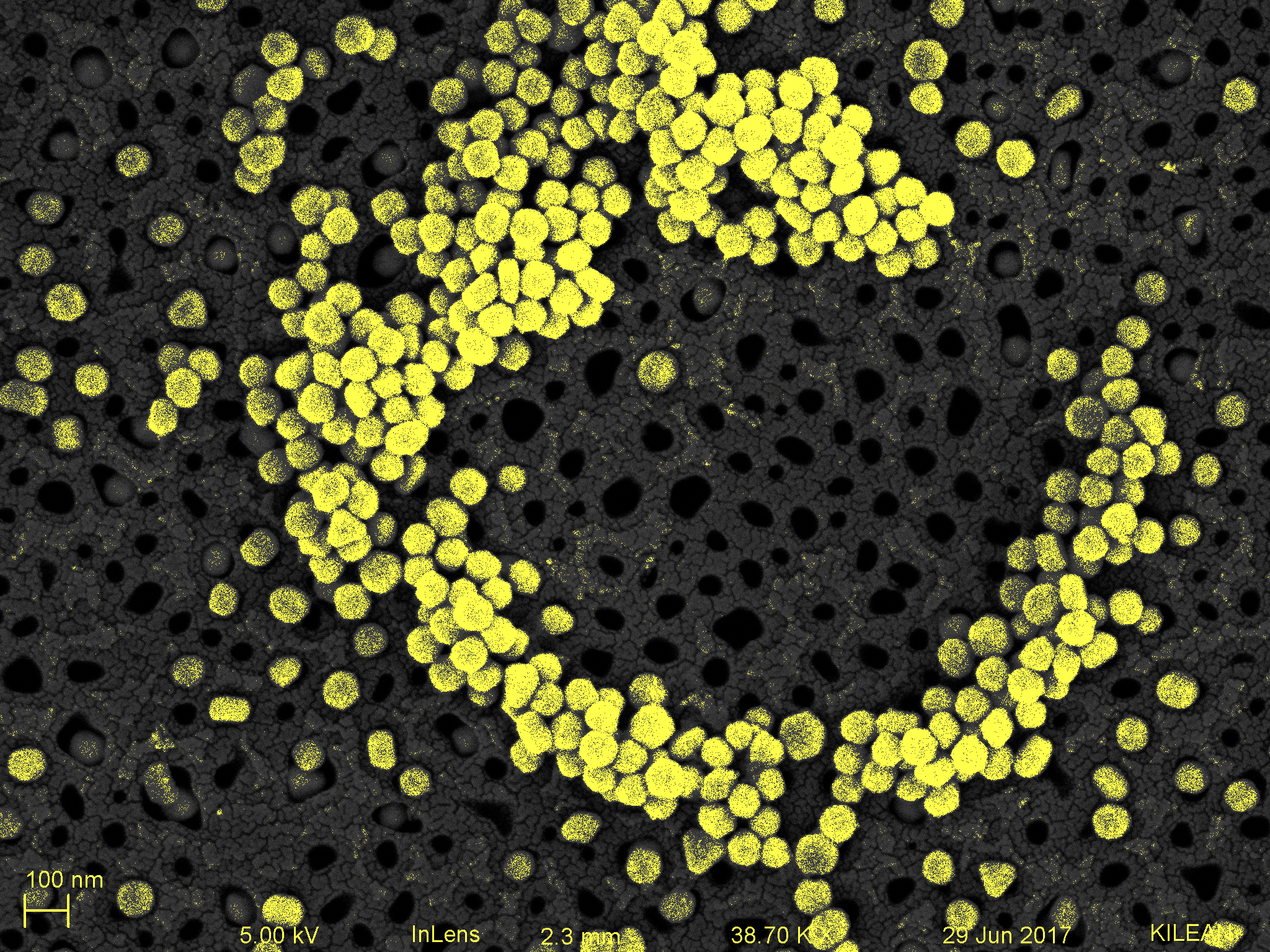
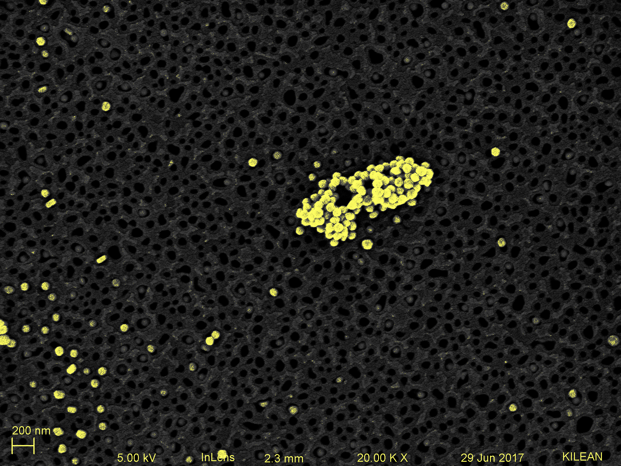
Within the past few weeks we have been working on finalizing an architecture for PETL dialysis that can be cut out, assembled, and disassembled repetitively with precision. Within the past week we were able to replicate the results Kilean achieved back in March with capturing gold particles in the membrane using tangential two-pump flow. 80 nm gold particles were caught in the membrane indicating that there was no leakage around the chip seal and the through-flow was indeed through the actual membrane. Given that these results can be replicated, our next step is to attempt filtering viruses out of solution to image at high resolution.
~Anthony