Transwell Edge Effects – Part 2
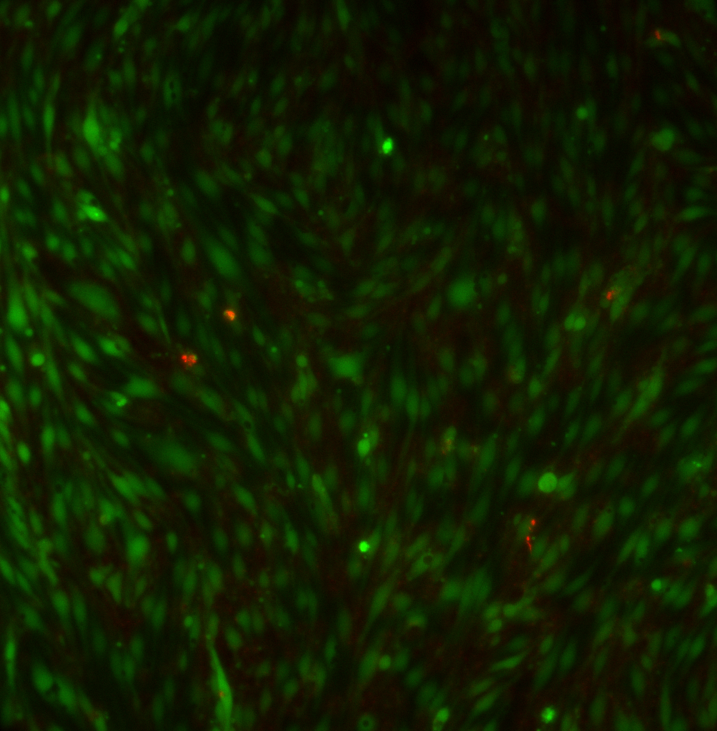
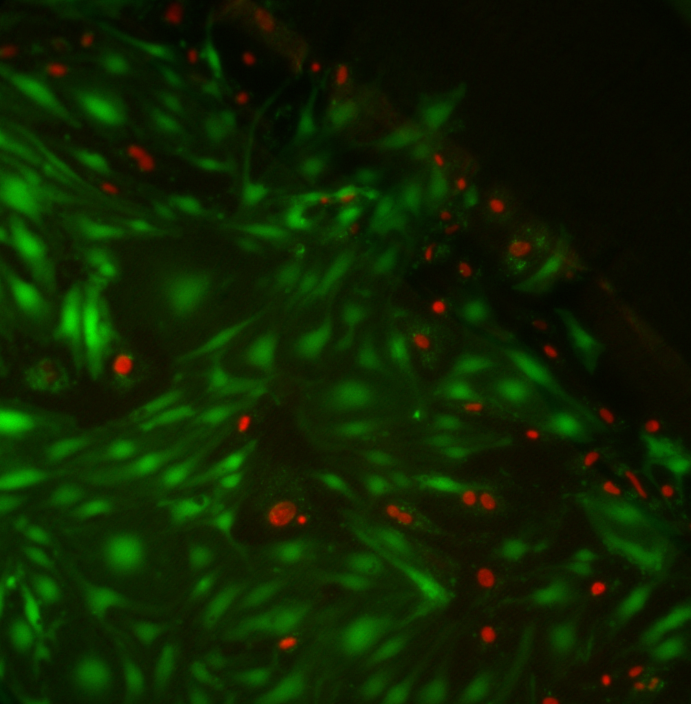
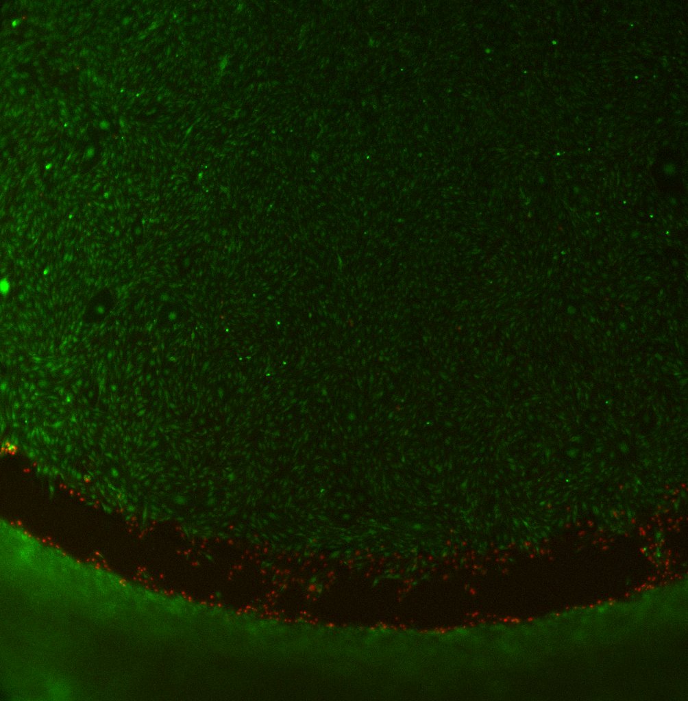
A while ago, I posted some images of cells grown on commercial polyester (PET) transwells. Here’s the post. I found that there was an annulus of dead cells around the edge of the transwell when cells were grown on the bottom of the membrane. I contacted Corning and they said that these dead cells were attached to the part of the membrane that was ultrasonically welded to the supporting PS material. This doesn’t make any sense to me, so I redid the experiment. These are P8 bEnd3 cells seeded on the transwells @ 100000 cells/cm2 and grown well past confluence to 7 days. Cells stained with Live/Dead assay. Images at 20X, unless otherwise noted.
Middle of PET membrane shows confluent endothelial monolayer with few dead cells:
At the edge, however, there are lots of dead cells. The edge of the membrane is toward the right corner of this image:
At 4X, this effect is even more obvious:
The edge effects are less pronounced when cells are grown on top of the PET. Here, the edge is the bright green thing in the bottom left:
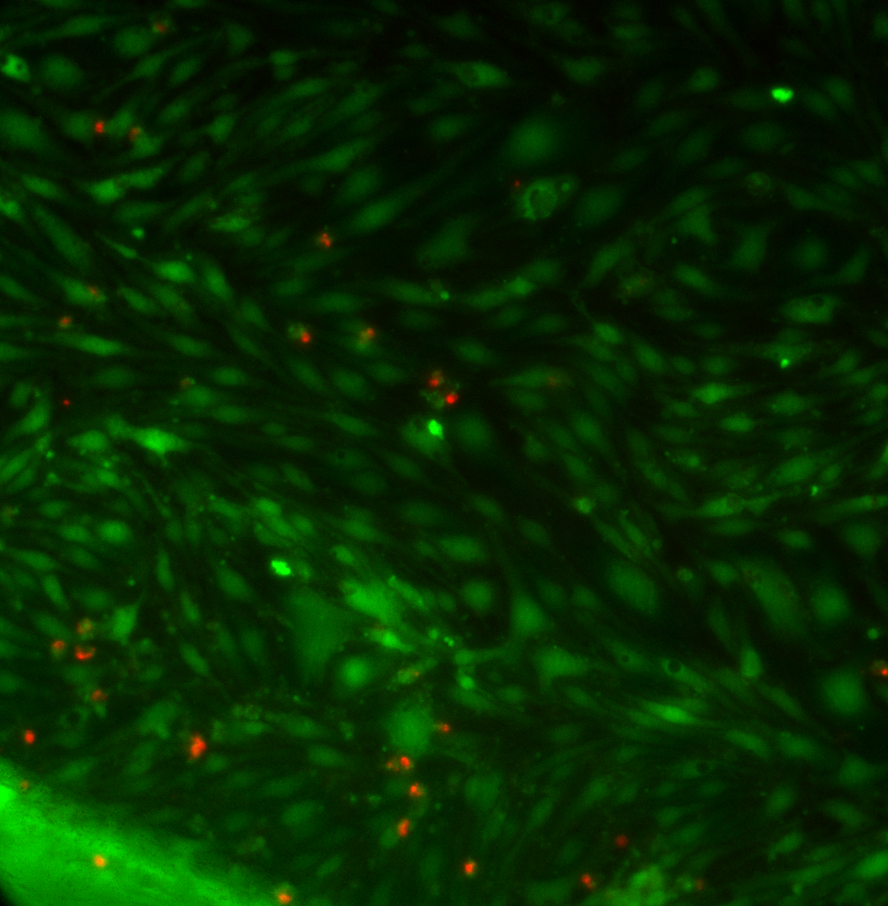 This confirms the previous experiment. Since the endothelial cells are the barrier cells, it is important to keep them as ‘happy’ as possible. I’m going to switch to growing them on the top of PET transwells.
For pnc-Si, this is not an issue since the active membrane area is far from the edges of the Sepcon plastic.