BBB co-culture (2)
In my last post (the first co-culture), I showed that bEnd3 cells exhibited a different morphology when they were co-cultured. I also decided that I would reverse the orientation of the co-culture in order to better visualize the endothelial barrier. That is, grow bEnd3 on the basolateral side and NG10815 on the apical side of pnc-Si transwells. This post outlines these results.
Here, I seeded 50000 P13 bEnd3 cells/cm2 on the bottom of pnc-Si and PET membranes, let them attach for 2 hours, inverted the transwells into a 24-well plate and then seeded 25000 P17 NG10815 cells/cm2 into the apical well. This is half the density of NG108-15 that I used last time. I tracked TEER and imaged after 7 days.
bEnd3 alone shows not as many vacuoles as usual, but typical morphology:

Coculture:
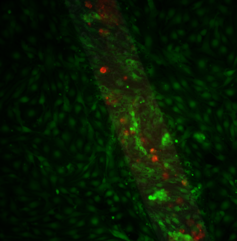
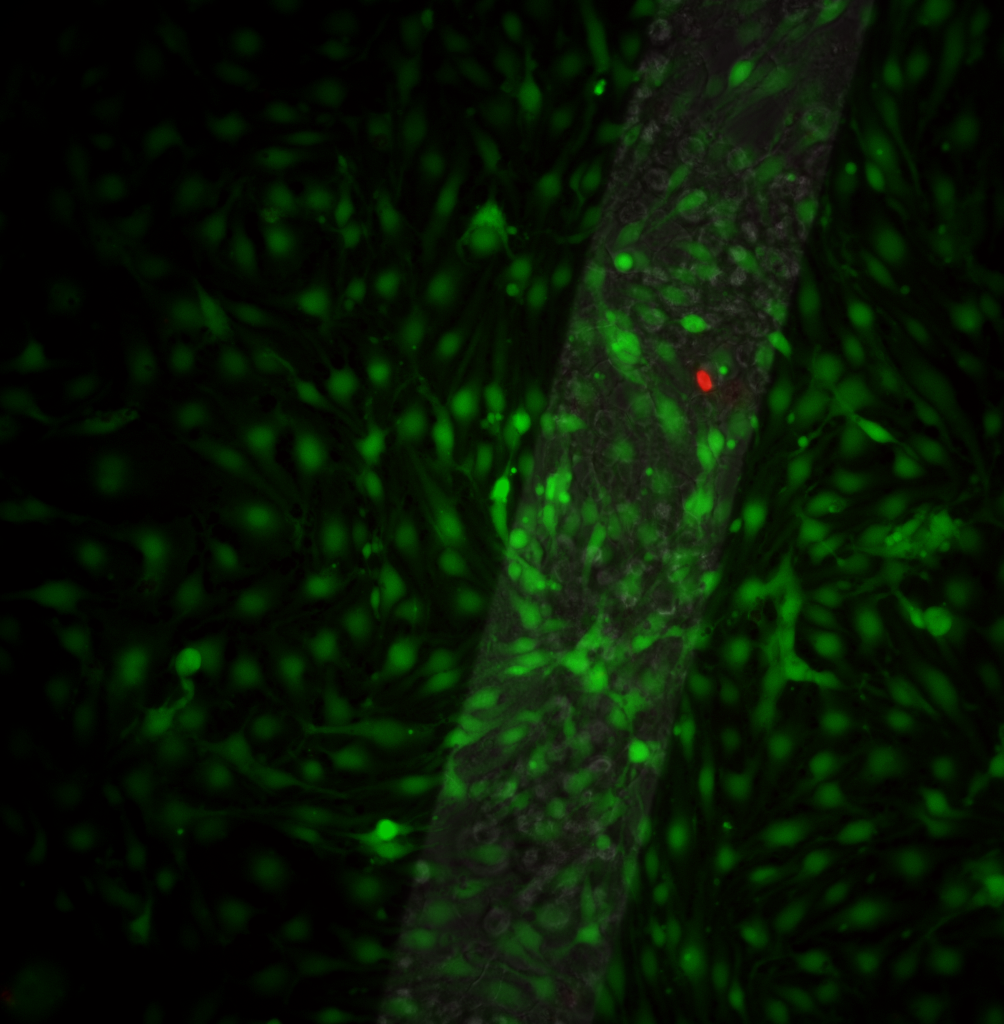
These are images from 2 different transwells – I stained the bEnd3 side only. You can see different morphologies – the top sample had endothelial cells with many vacuoles. The bottom sample had very few vacuoles. Many of the NG108-15 cells were dead on the top sample. If NG10815 multilayers act to choke off permeability, could the presence of dead cells increase permeability and thus allow enhanced vacuole formation? I kind of doubt it but I’m not sure what else could explain this difference.