Three different thicknesses membranes from backside
This is my (Jim) summary of the results thus far for NPN vs NPN-O and Front Side/Back side comparisons. More to come …  *************************************************************************************************************
As thesis indicated, the thicker membrane is, the larger average pore size will be. From my process, the average pore size for 50nm membrane is between 9nm and 10nm; the roundness (how circular the pores are) is around 0.74, which means the minor axis of the pores are 0.74*the major axis of the pores. The average pore size for 75nm membrane is between 19nm and 20nm; the roundness is about 0.73. The average pore size for 100nm membrane is between 36nm and 37nm; the roundness is about 0.71.
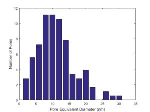
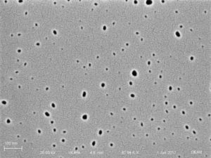
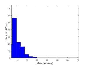
Image1-1(up): Representative of 50nm membrane pore size distribution.
Image1-2(middle): Image of the pores under 97.94k magnification.
Image1-3(down): Minor axis histogram of 50nm membrane pores.
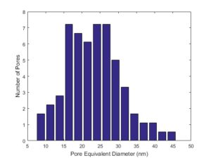
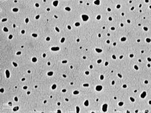

Image2-1(up): Representative of 75nm membrane pore size distribution.
Image2-2(middle): Image of the pores under 100k magnification.
Image2-3(down): Minor axis histogram of 75nm membrane pores.
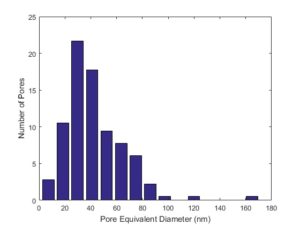
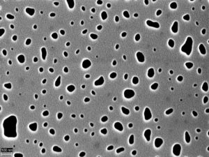
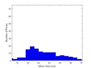
Image3-1(up): Representative of 100nm membrane pore size distribution.
Image3-2(middle): Image of pores (I don’t know the magnification for this image, it should be smaller than 100k).
Image3-3(down): Minor axis histogram of 100nm membrane pores.
| p(porosity) | r (average radius) (nm) | Lp-p(average distance between pores) (nm) | ||||
| 50nm | 0.01606 | 4.74815 | 71.3588 | |||
| 75nm | 0.0593 | 10.4645 | 81.8466 | |||
| 100nm | 0.09334 | 18.2499 | 113.772 | |||
Table1: this table includes the average porosity, average pore radius and average distance between pores for all three thickness membranes.
We can clearly see that pores are getting larger. For three thicknesses membranes, they can capture three different particle size ranges.
The reason why I look from backside is that backside pores are smaller than the frontside pores. The backside pores finally decide how much the blood can flow through the pores. And backside pores are easier to identify and measure.
I went to the SEM laboratory on June 5th with Dean to grab some images from 50nm membranes. Other than typical pore images, we found something interesting on the edge of each membrane section.
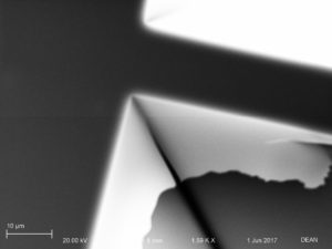
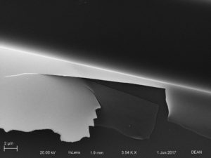
We are confused about what it is and we could not focus on it close enough to see if there are pores on it. On my opinion, it might be an edge scratch along with the manufacture of membrane.
PS. I have updated all the magnifications of all the images. I also added a table contains other important information.
Nice work JK. Can you please update this post to include minor axis histograms since that is the selective dimension. Also include some measure of how circular the pores are for each material.
Just updated!
Which 50nm SiN was this? The small pores (Lot 1211) or the standard pores (any other lot)? Also, this image was not at 100kX mag. I am not sure that it makes much difference, but technically the pores are slightly larger than listed.
The item in the last images is the backside mask. This flap of SiN is non-porous and due to undercutting of the backside mask during the bulk etch. All chips have this to varying degrees depending on the size and shape of the frontside membranes.