Cells on squares vs. slits
Last week, I finished up an initial experiment to investigate whether pnc-Si membrane geometry affects cell morphology. To do this, I used a mask with 2×2 arrays of different size squares and also samples with 4 slits of different lengths and widths. A lot of the samples broke before I got the wafer, so this is the only experiment I got from this wafer. I was hoping to find a magic aspect ratio that caused cells to form tubes.
These were P13 cells, seeded at 50000 cells/cm2 and grown for 7 days before staining with Live/Dead. All of the longest slits except 1 broke during culture or feeding (these were ~ 4mm long), regardless of their width.
Here are the images from the square window samples. Only 2 of the 4 designs I made cleared the etch, so I only had 2×2 arrays of ~425 and 200um2 membranes:
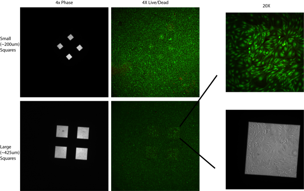
There is no change in cell morphology between cells on free-standing vs. supported pnc-Si (I included phase images to show where the windows are). The rightmost panels show 20X images of cells on 1 of the large squares – vacuoles are present.
The slits are ~200um by 4400, 2000, 1000 and 500um (so aspect ratios of ~22, 10, 5 and 2.5). The top panels are 4X, bottom is 20X.
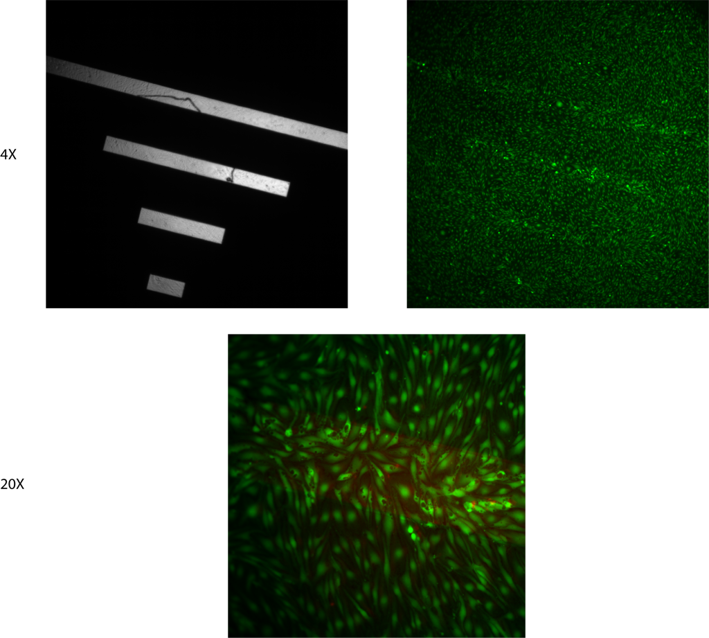 Like I mentioned earlier, I only had 1 sample survive with all 4 slits attached, so these are the only images of cells on membrane slits. There is no tube formation over the free-standing membranes, although there are vacuoles present, as expected. I’d say this is inconclusive, so I redesigned the mask in order to repeat this experiment.