Blood-brain barrier co-culture (trial 4)
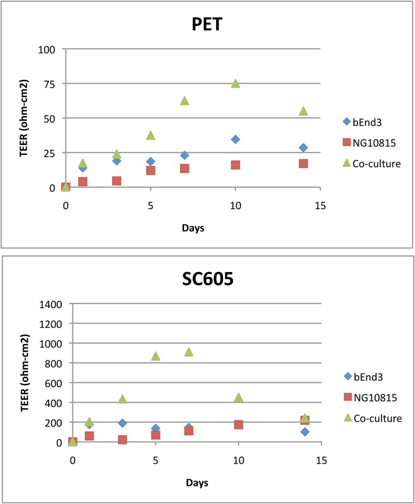
This is simply the latest repeat of a blood-brain barrier co-culture on PET and pnc-Si transwells. These are P17 bEnd and P24 NG10815, each seeded at 50000 cells/cm2. As usual, I seeded bEnd on the pnc-Si side, upside down, and NG10815 on the well side, facing up. I took TEER measurements every other day for a week and then a couple more times out to 14 days, after which I stopped the culture and stained the cells with Live/Dead.
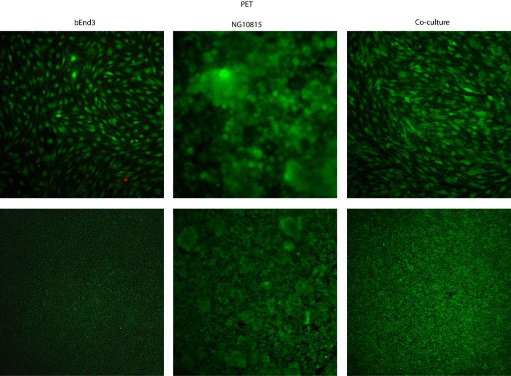
On PET (top rows 20X, bottom rows 4X), cells look ‘normal’. The bEnd cells both for the mono- and co-cultures had similar morphologies and there were scattered vacuoles. NG10815 grew as clumps. I included 4X images to show the lack of tube formation in these samples.
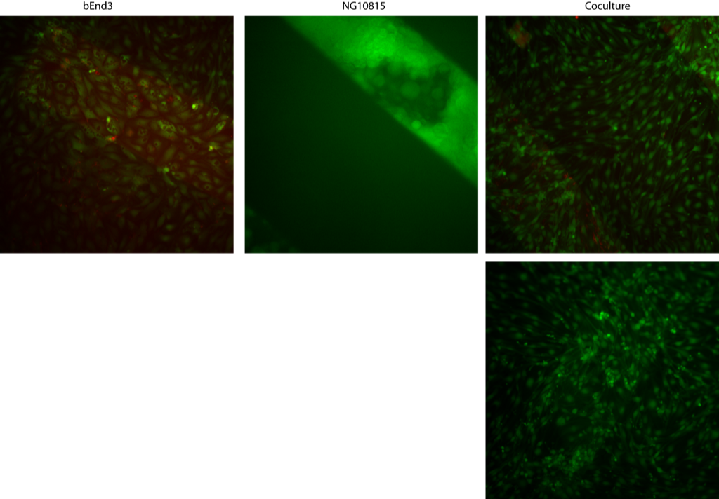
On pnc-Si, the bEnd alone had lots of vacuoles and the NG10815 grew as clumps (the high background for NG10815 is from fluorescence in the apical well, which I didn’t want to aspirate for fear of shearing off these loosely adherent cells). I included 2 images of bEnd from co-cultures (right column) – both show very little vacuole formation in bEnd3 cells when co-cultured with NG108-15. I’ve seen this before here, here partly, but not here although these cells are forming tubes. It seems like the presence of NG10815 on the well-side of pnc-Si transwells might act as a blockage of permeability, which inhibits vacuole formation as if those bEnd were on an impermeable floor.
Here are the TEER data, which are similar to these and not terribly different from these:
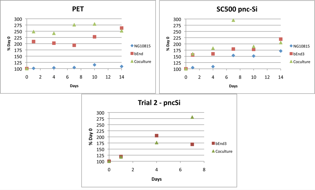
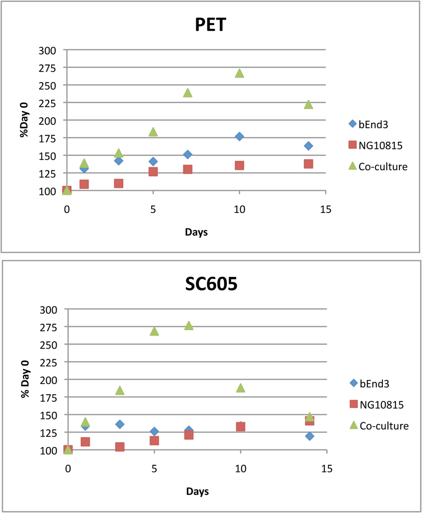
There is a gradual (and eventually super-additive) increase in TEER for the co-culture on PET, which then falls at day 14. There is a rapid and immediately super-additive increase in TEER for the co-culture on pnc-Si, which then falls slowly over the next week. This is a prolonged TEER spike. For pnc-Si, the bEnd TEER alone looks terrible and is actually overtaken by NG10815 at late times. I’m not sure about this since the bEnd3 cells alone looked fine and were viable after 14 days (unless the vacuoles have a role in increasing trans-endothelial permeability here). So, in my 4 BBB experiments to date, I’ve seen a spike in TEER for co-cultures on pnc-Si every time (I’m being optimistic and assuming that this experiment and trial 2 simply didn’t last long enough to see the downward trend) and occasionally spikes on PET. TEER tends to increase faster on pnc-Si, as well. I think it might be interesting to do a time course with a CellTracker stain to track vacuole formation/viability with TEER measurements.
UPDATE(MAY 17, 2010): TEER Data as “Net TEER”