Possible Gel-Box Outgassing on Porous SiO2 Membranes
In previous experiments attempting to culture ADSC on non-porous and porous (0.5 µm, 3.0 µm) SiO2 membranes, we were having difficulty getting the ADSC to attach to the membranes. A majority of the ADSC did not appear well-spread. While in culture or staining, ADSC would detach from the membrane with little to no agitation. Changes to the Geltrex-coating protocol and the staining protocol did not appear to make a difference.
These membranes had been popped out of wafer lot 1110 and stored in Gel-Pak gel boxes for approximately (4) months. Tom is hypothesizing that the gel boxes may have outgassed and deposited on the membranes, leading to difficulty in cell attachment. The purpose of this post is to provide images of these “dirty” membranes and attempts at cleaning them. All membranes were sputter-coated with gold for 50 seconds. All images were acquired at a working distance of 9.6 mm.
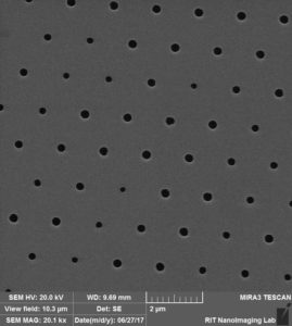
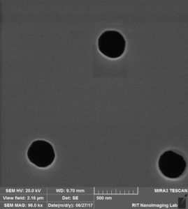
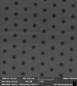
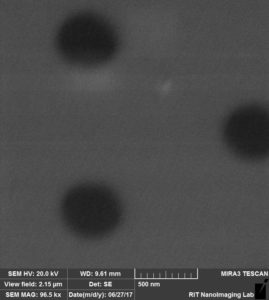
SEM images of a “clean” control membrane. This 0.5 µm SiO2 membrane had been popped out of a wafer and stored in a 24-well plate for approximately (4) months. he images, taken at 10, 20, and 96 kx, are very clear and are of the expected imaging quality. Interestingly, yet not entirely related to this project, there appears to be some occluded pores and variation in pore diameter across this sample.
 |
 |
 |
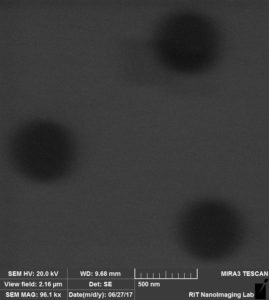
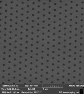
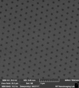
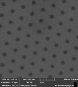
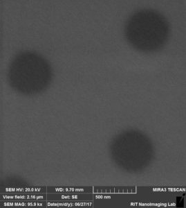
SEM images of a “dirty” control membrane. This 0.5 µm SiO2 membrane had been popped out of a wafer and stored in a gel box for approximately (4) months. The images, also taken at 10, 20, and 96 kx are fuzzy and unfocused. We were unable to take clear images of these membranes. Dr. Hailstone suggested that this inability to acquire clear images was due to some sort of residue that has deposited on the surface.
 |
 |
 |
We attempted to clean the membranes using a Plasma Cleaner (Harrick Plasma, PDC-32G). The sample shown below was plasma cleaned at medium power for 2 minutes. As you can see, no improvement was made to the imaging quality.
 |
 |
 |
The sample shown below was plasma cleaned at high power for 10 minutes. Similarly, no improvement was made to the imaging quality.
 |
 |
 |
Given that the plasma cleaning did not improve the imaging quality of these membranes, we are assuming that the membranes are still coated with residue. We would like to discuss other ideas to clean the membranes.
Thank you to Aslan for his assistance with everything.
I’m sorry to hear that this may be causing cell attachment issues. I think Greg M and Kevin W showed by SIMS that this was happening a couple of years back. SiMPOre’s TEM windows customers have reported similar possible coatings when trying to deposit highly polar samples on older lots.
Yes we had some big problems with what looked like uncured PDMS migration onto the surface, which led to problems with cell death and poor adhesion. Since we shifted to Greg’s 3D printed cartridge boxes we haven’t had this issue. Clean-up probably requires something fairly aggressive solvent-wise, directed at solubilising the elastomer. There are various options but none of them are pretty.
Question for next NRG discussion: have we seen any evidence of this on NPN or microporous SiN materials? They would have to have been stored in gel boxes for a long time. So far I think we’ve seen it on pnc-Si, MgF2, and oxide membranes, but not SiN. It matters because the gel boxes are handy and cheap, if they can be used.