SEM Images of Barcikowski Samples
With Joe’s help, I was able to take some SEM images of the samples that I performed my previous set of separation experiments on.
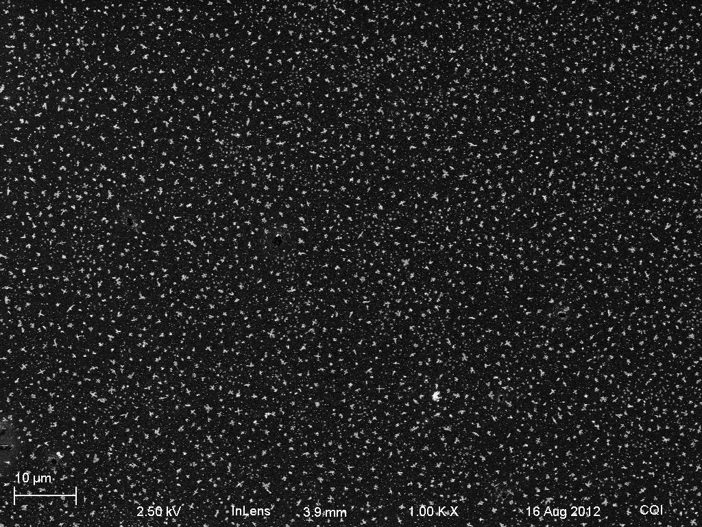
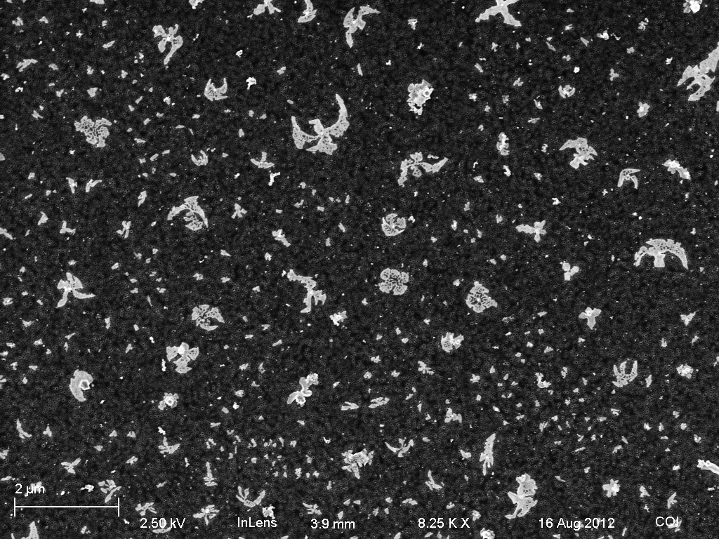
Sample 1: The obvious aggregation in this sample (exemplified by the ‘victory eagle’ blot in the second image) is observed in all five of the samples. What we can’t know is the extent to which this clumping is a result of drying out the samples, as opposed to the aggregation that is occuring in solution. Notice in the third picture that there are some lonely gold particles that did not clump.

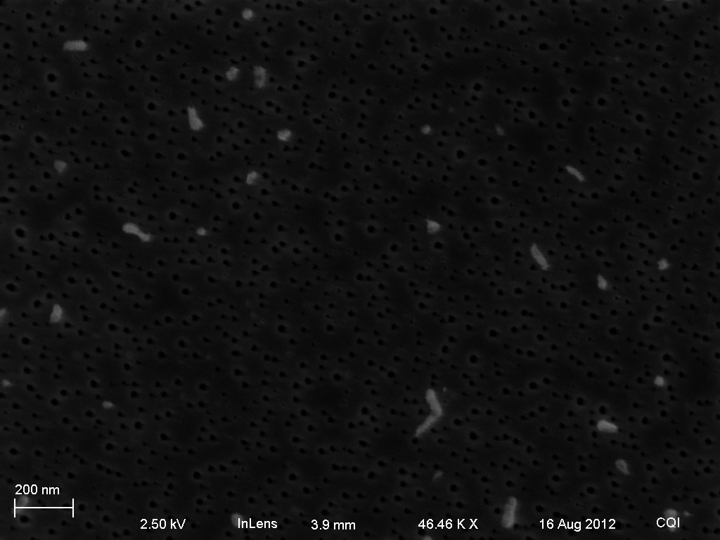
Sample 2: Although we still see clumping, this sample seems to be at a lower concentration. The second picture (and to some extent all of the pictures) is blurry because of the native oxide layer on the silicon – the sample was dried onto a non-freestanding pnc-Si membrane


Sample 3:

Sample 4:


Platinum:

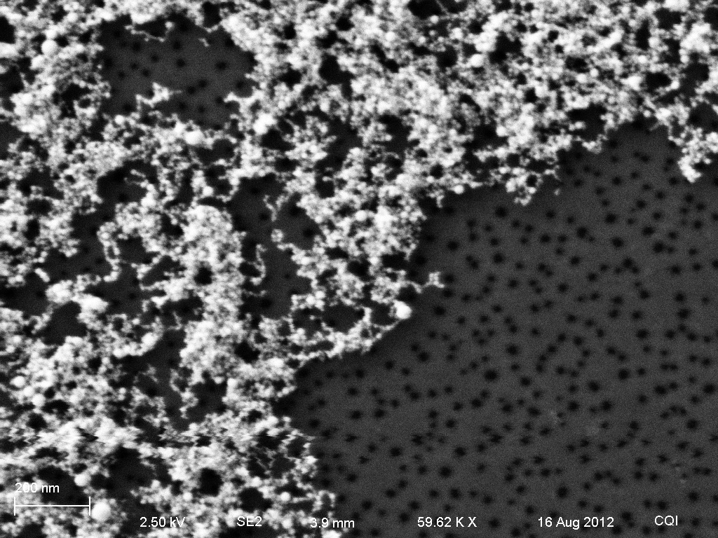

So in each picture, we see both clumps and individual particles of a size we want.