TEM images of actin filaments on Formvar grid
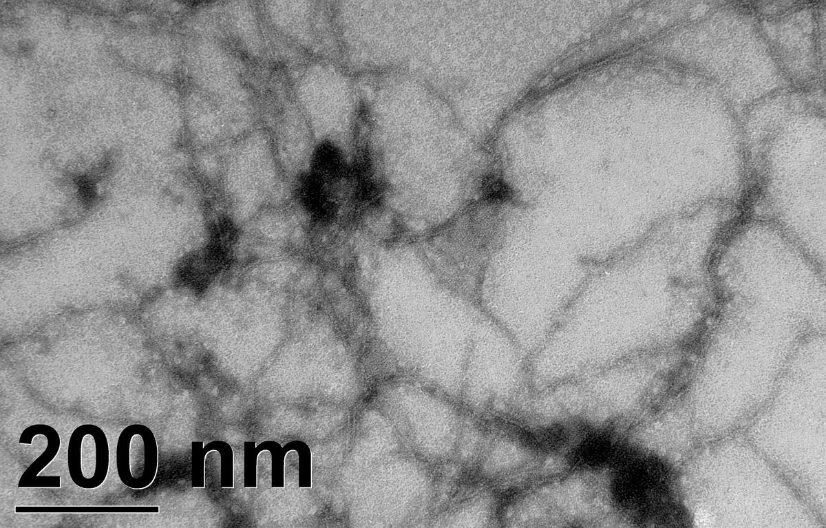
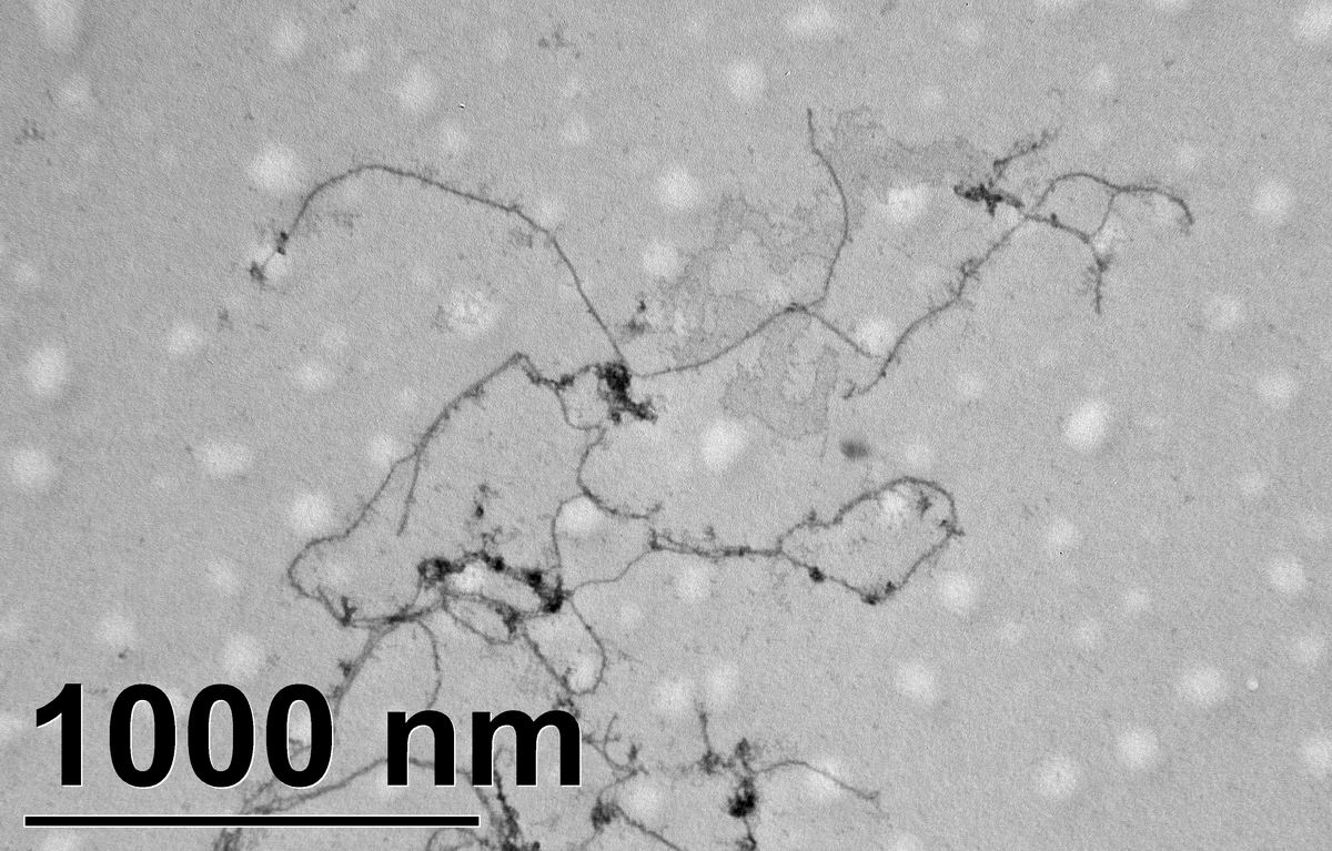 Here are images of actin filaments on Formvar coated 200 mesh nickel grid. Actin filaments were polymerized exactly as before.
 TEM sample preparation procedure is:
1. add actin solution on Formvar grid and wait 90 sec
2. remove liquid, add same volume of 2% UA and wait 90 sec
3. remove liquid and load on TEM sample loader
What are the differences comparing to our nanomembranes?
1. actin filaments did’t clump on Formvar grid
2. actin filaments picked up stain better on Formvar grid than they did on nanomembranes
3. we saw more filaments on Formvar grid(maybe not…)
What does this mean?
1. Different surface charges??
2. Different adhesion ability to proteins(Jim)*
3. Other…
*: Seems Formvar does have good adhesion. “Wolff AM. The use of carbon-coated formvar films as bacterial adhesion substrates for scanning electron microscopy. J Electron Microsc Tech. 1988 Nov;10(3):315-6”
we could not focus images at high mag(also low mag but not obvious)because of the new equipment is malfunctioned.