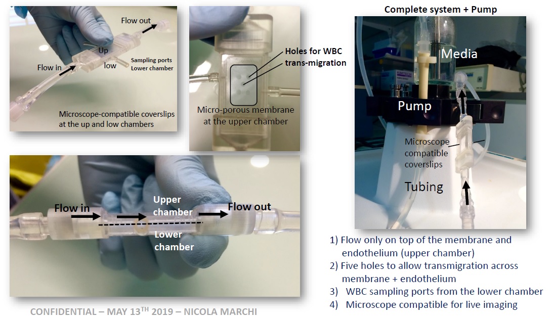
Modeling leukocyte transmigration thorough a blood-brain barrier model
This post is dedicated to our ongoing collaboration with Dr. Marchi at Centre national de la recherche scientifique (CNRS) in France. The basic concept of the project is simple (Fig.1) and involves attaching a microporous membrane on top a 3D printed “drawer”, seeding endothelial cells on top to confluency, seeding epithelial cells on the bottom to confluency, and allowing potential leukocyte transmigration through endothelium and microporous membrane.
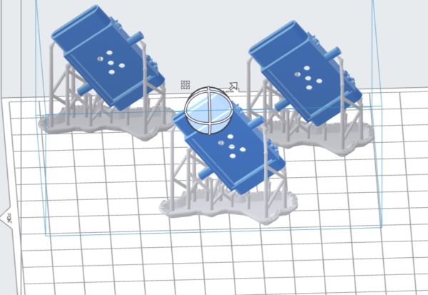
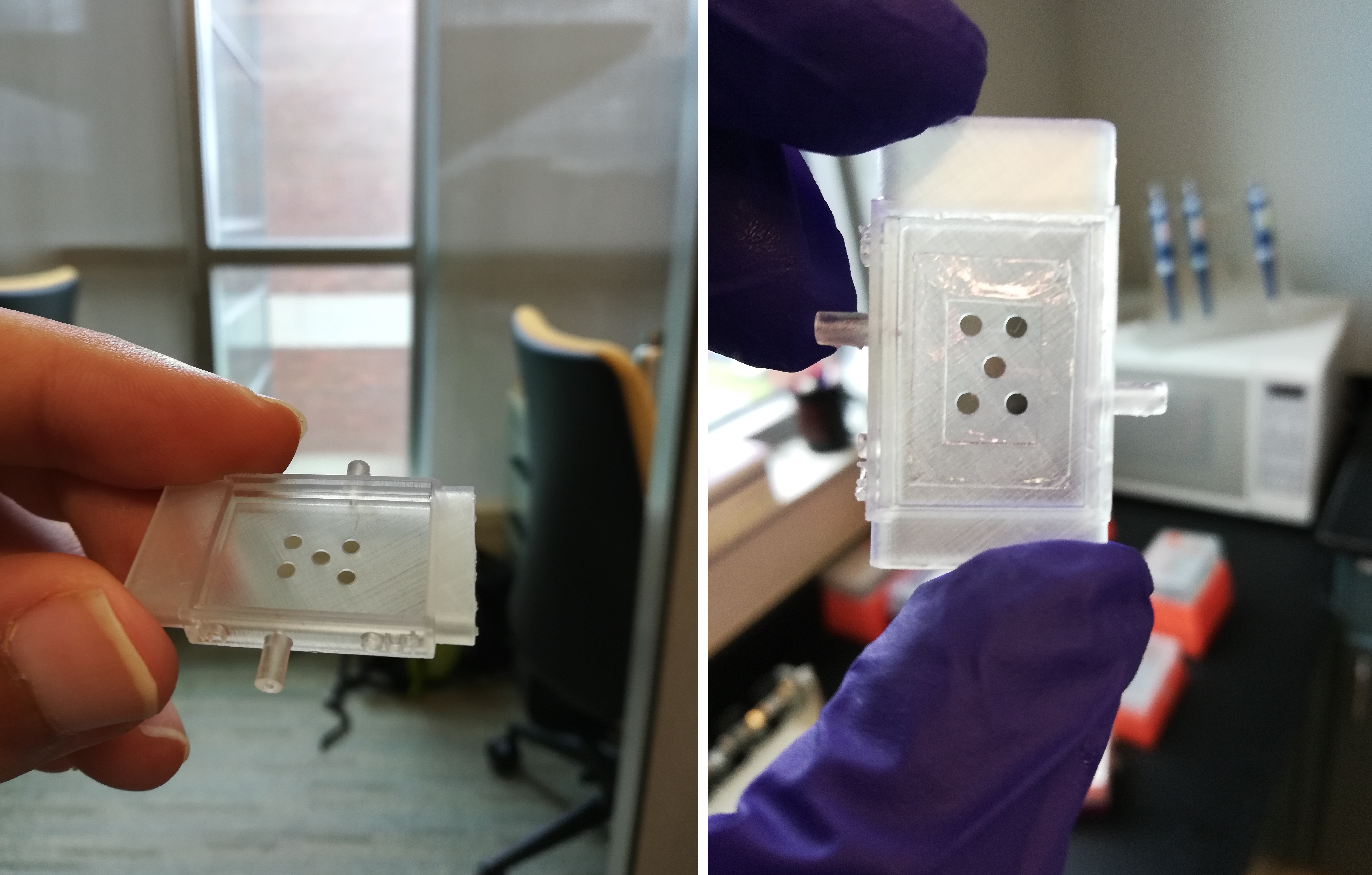
Initially, we would get the 3D printed samples from France, attach membranes to them and send them back for the initial experiments. After some shipping issues, we tried making the drawers and had some problems due to the very small thickness of the 3D printed sample in the middle and we were able to resolve that problem.
Next step was to attach membranes to these drawers. There were several considerations, most importantly that the membrane should be attached without making any disturbance in the media flow on top of the membrane. For this purpose, we used epoxy glue. We did extensive cytotoxicity assays for the drawers and the glue (ISO 10993-5) and found no significant effect.
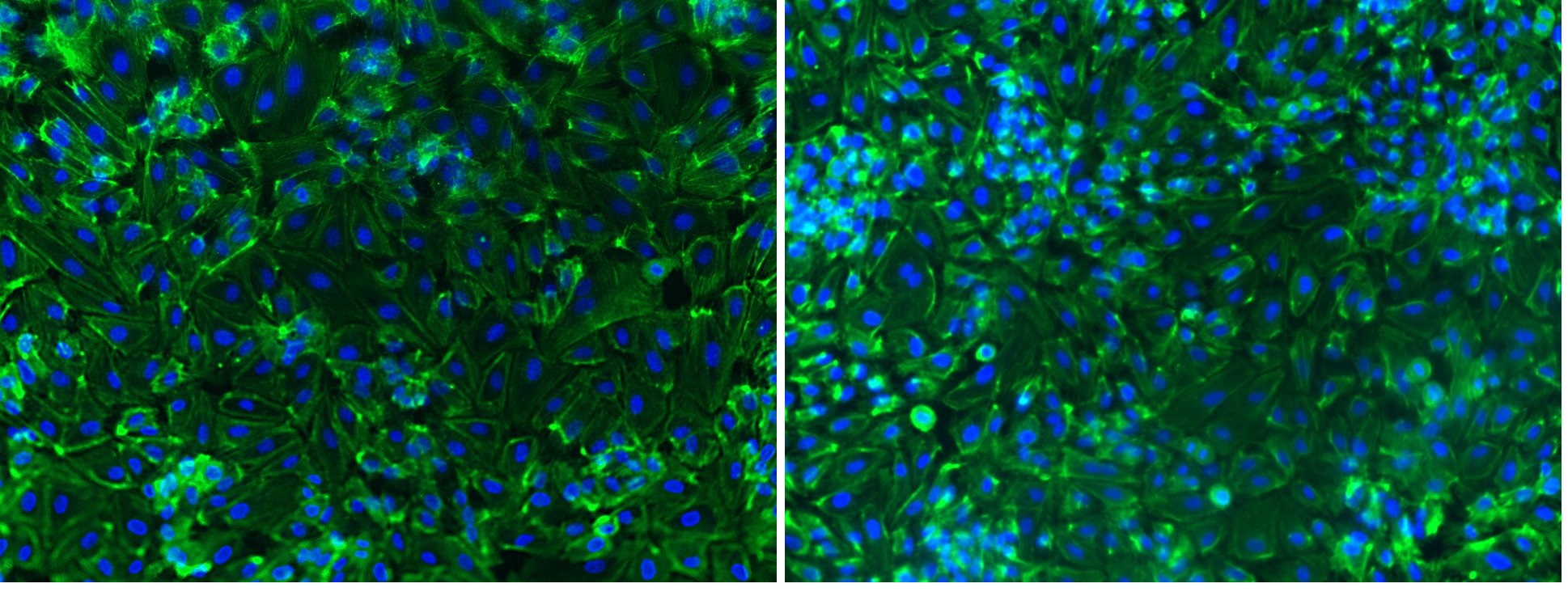
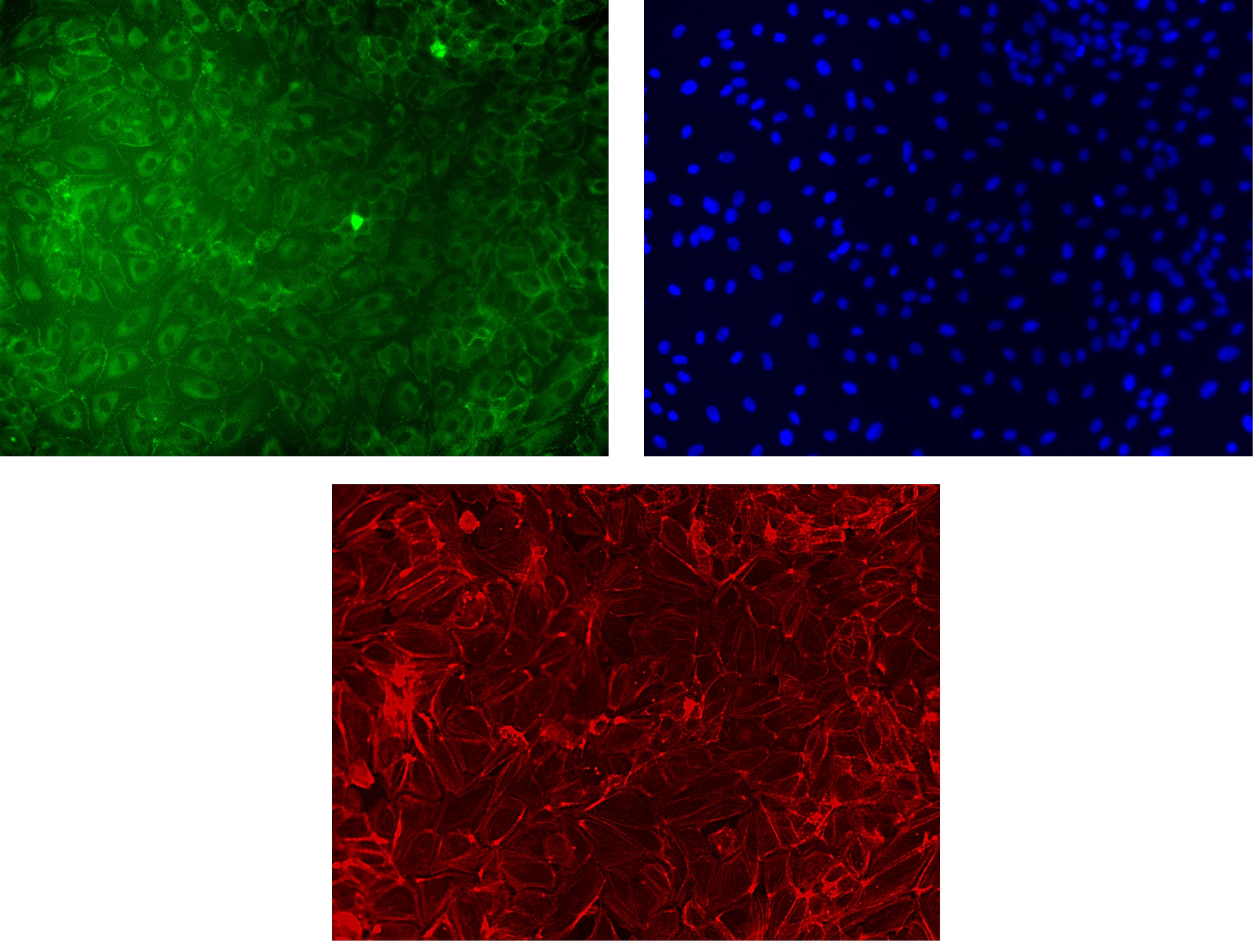
The next step was to make sure that the membranes possessed proper characteristics for enabling a proper endothelial barrier formation without the necessity of using artificial over-seeding. The following samples were seeded at a 10k/cm2 density of HUVECs and imaged after 72 hrs. No treatment and/or incubation with media was used.
The next step is to send samples with different drawer design to France. Hopefully, there is not much left on the fabrication/membrane characterization front for this project.