Discoloration – autoclave #2
I repeated the autoclave experiment from earlier this week, but this time I also used the Sepcon format. I used wafer 662, which was contaminated with large spots. This was done in the oven in RPMI-1640 with FBS (no RTP). I think my 0 hour pictures were accidentally erased from the camera, but I think everyone knows what day 0 pictures typically look like.
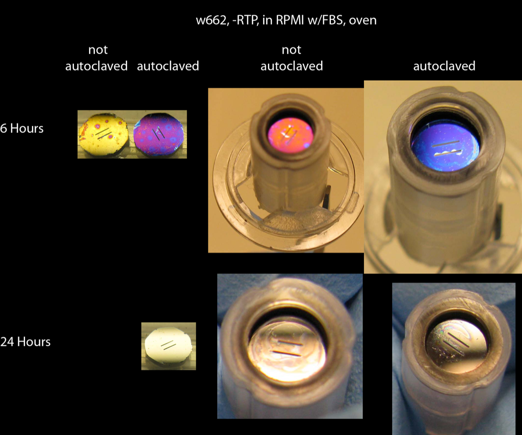
Discoloration was faster in this experiment due to the use of cell media and not NaCl. As before, the autoclaved chip discolored slower than the chip that was not autoclaved. The trend was the same in the Sepcon format. The Sepcons discolored much faster than in my previous post. This is partly due to the incubator vs. oven effect. Also, these membranes were broken, so there was more transport between the apical and basolateral wells of the Sepcon (the 2 sides weren’t really isolated). My previous Sepcon post employed blank chips, so the 2 sides were truly separated.
I removed the chip from the autoclaved Sepcon sample and took a picture:
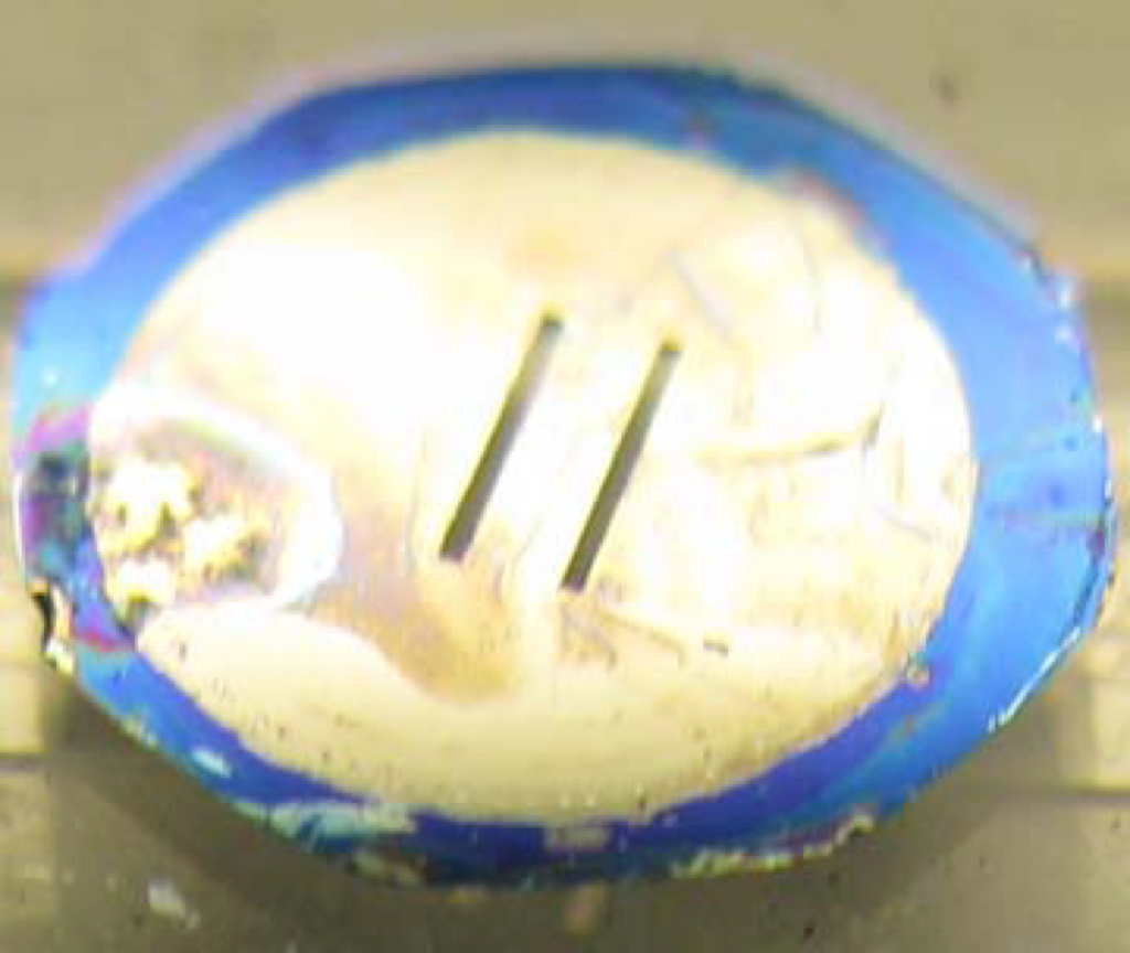
Cool looking but not surprising – you can clearly see where the O-ring was squeezed against the pnc-Si in the Sepcon format.
I did see something like the last image using cloning rings. Also, my post on discoloration in Sepcon has similar colored O-ring.