Adipose Stem Cells on Nitride LiftOff Membranes (Part 2)
Background: In a prior post I showed that we had seeded cells on both nano- and micro-porous SiN Liftoff Membranes. The nanoporous proved to be rather weak and showed numerous broken windows after seeding the cells.
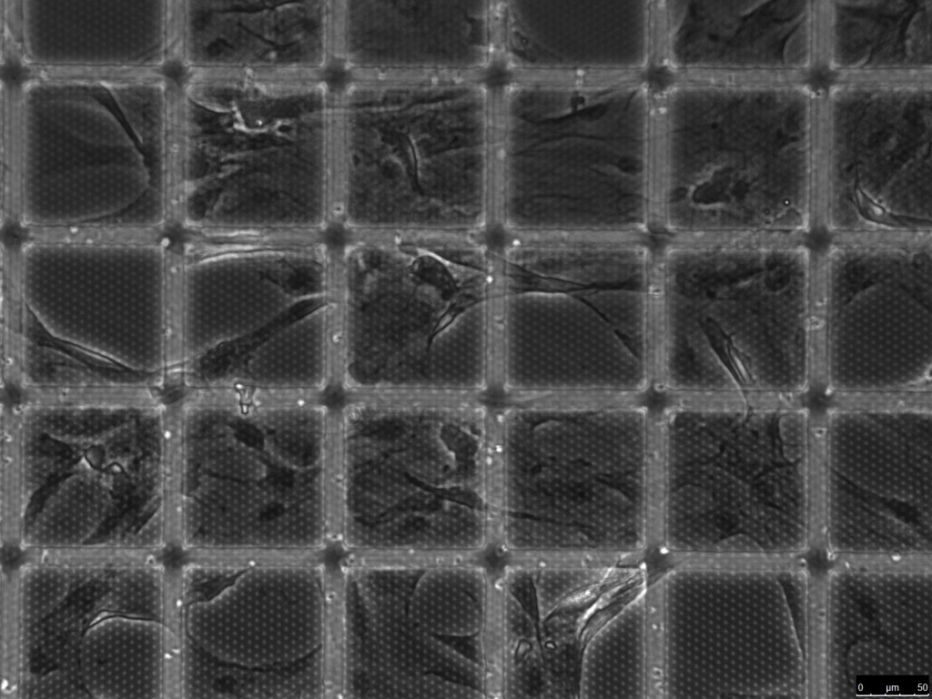
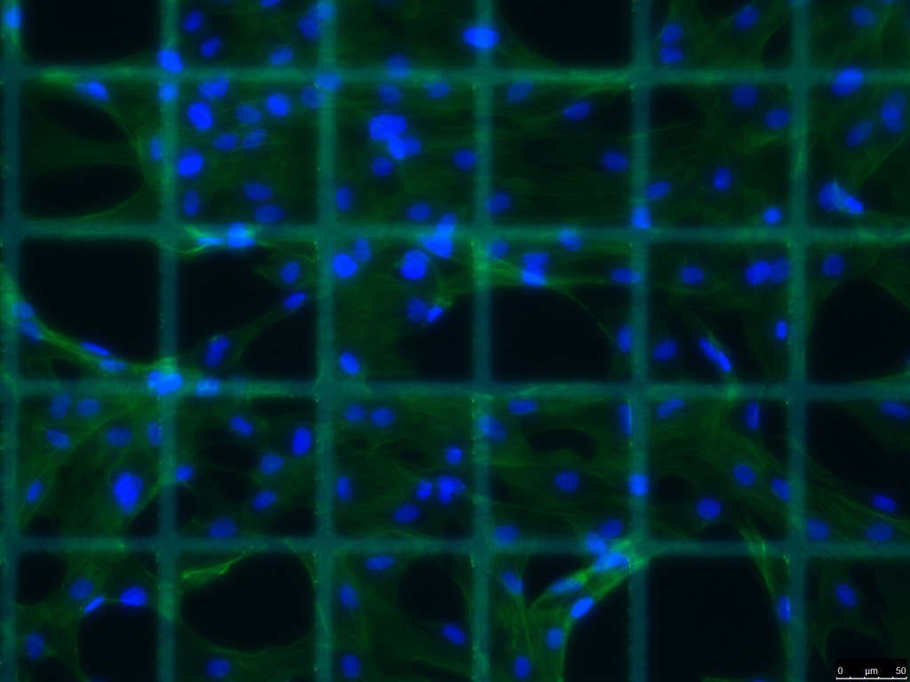
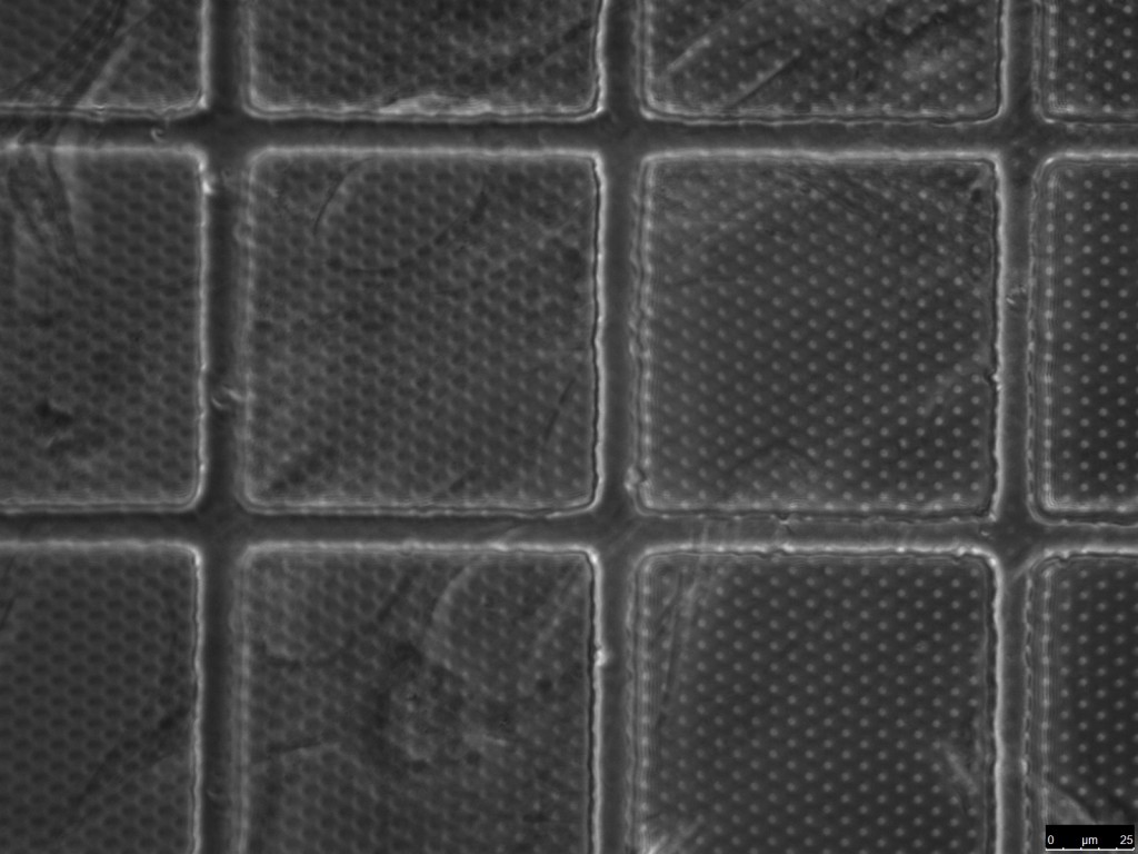
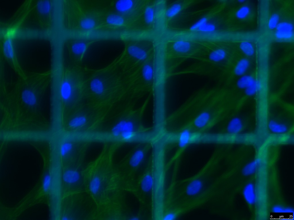
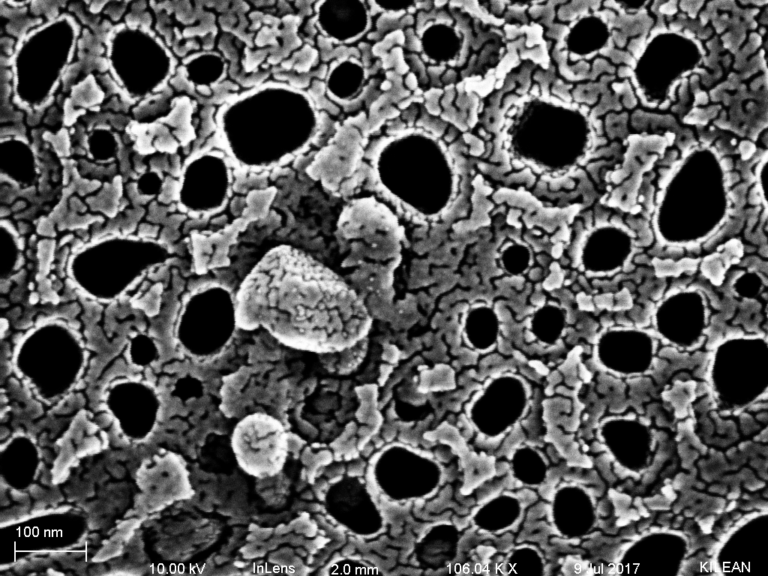
Summary/Conclusions: In this follow up post, I seeded adipose derived stem cells on 50 nm thick microporous SiN liftoff membranes with 100 x 100 micron windows in the SU-8 scaffold. These membranes saw an incredible amount of handling and manipulation with minimal window failure or tearing. The membranes sat for somewhere between 6-12 months on the shelf in a petri dish prior to our use. Bob attempted to bond silicone gaskets to the top side of the membrane and added liquid PDMS to help adhere these gaskets which were designed to retain the cells. I washed the device in 50% ethanol to sterilize. From pre-incubation to cell seeding, fixation, permeabilization, staining and washing, these membranes saw 14 liquid exchange steps/procedures. The most striking detail beyond the stability is that the 50 nm SiN membranes did not interfere with inverted fluorescence imaging of cells on the top of the membrane. We found previously with 120 nm SiN that the pores could be identified in fluorescence images by a brighter signal.
Methods: The liftoff membrane was suspended above a glass coverslip with a 1 mm thick silicone gasket. The membrane was incubated with a 1% w/v basement membrane protein mixture (Geltrex) in 1x PBS for 30 minutes at room temperature and then washed prior to seeding cells. ADSC cells (passage 3) were cultured in non-differentiating proliferation media for 4 days on the top side of the membrane. Cells were stained with DAPI (blue) and Fluoroscein-Phalloidin (green). Images were collected on an inverted fluorescence microscope in 10x, 20x and 40x.
Pictures: