Neutrophil-endothelial transmigration through ECM gel scaffolded on microporous silicon nitride membrane
I have been working on developing a microvessel mimetic using our silicon membranes. Just as a recap of vascular biology, the basic components of a microvessel are endothelial cells, the basement membrane secreted by them (presence of perciytes is a plus), and extracellular matrix (ECM) on the abluminal side. This ECM is predominantly composed of type 1 collagen, although the exact composition of this matrix differ from tissue to tissue. I am trying to use our membrane to replicate this structure. I am using commercially available type 1 (rat-tail derived) collagen from here. The collagen, which is fibrous in nature, is dissolved into aqueous solutions by treating it with acid. The acidic pH helps to keep the collagen dissolved and in liquid state. In order to ‘gel’ the liquid, we need to raise the pH back to basic. The liquid solution sold online is 0.02 N acetic acid- a pretty weak acid. We need to add NaOH, PBS and water to make the solution basic, and isotonic. The recipe given online uses 1N NaOH, and 10x PBS, and dilutes each one of them by 10 times, i.e. 0.1 N sodium hydroxide and 1x PBS. Although isotonic, my pH calculations indicate that 0.1N is heavily basic and could be detrimental to cell growth. I tried to optimize the concentration by reducing the sodium hydroxide to be added without compromising gelling. The final recipe I used was:
For 100 ul of collagen gel:
(All components should be on ice. Pre-cool your vial before adding the collagen)
Step 0: Make sure your chip is good-to-go for cell seeding. Make sure that the Geltrex coating is all dried and everything is dry and clean.
Step 1: In a pre-chilled vial, add 28.8 ul of DI water
Step 2: Add 10 ul of 10X PBS
Step 3: Add 1.2 (yes!) ul of 1N NaOH (in DI water) . Ideal way is to make fresh solution every time. But if you want to spare the trouble, at least vortex the solution so that any undissolved/dried/precipitated sodium hydroxide will re-dissolve in water.
Step 4: (Make sure your surfaces are ready for gel addition since, collagen gels pretty quickly.) Add 60 ul of collagen in the mixture made above. At this stage, I used to sample 10 ul of this mixture and add on the pH strips just to get an idea about the resultant mixture.
Step 5: Using a long gel-loading tip, pipette ~10 ul of gel mixture in the trench behind our membrane. Spread it using similar tip so that you get a planar structure on the trench, and not a giant drop of viscous gel.
Step 6: Keep the chip in a closed, sterile and humidified petri-dish inside the incubator for 1 hour.
Step 7: After 1 hour your gel should be solidified. Try NOT to poke it with anything else you might drill a small hole in the gel.
Step 8: Pre-equillibriate the gel in cell media for at least 30-60 mins before actual experiment.
If you follow this protocol, the final gel concentration will be 3 mg/ml, with 1X PBS. Upon gelling it looks like below:
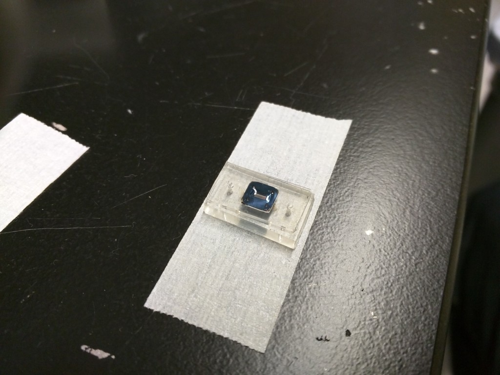
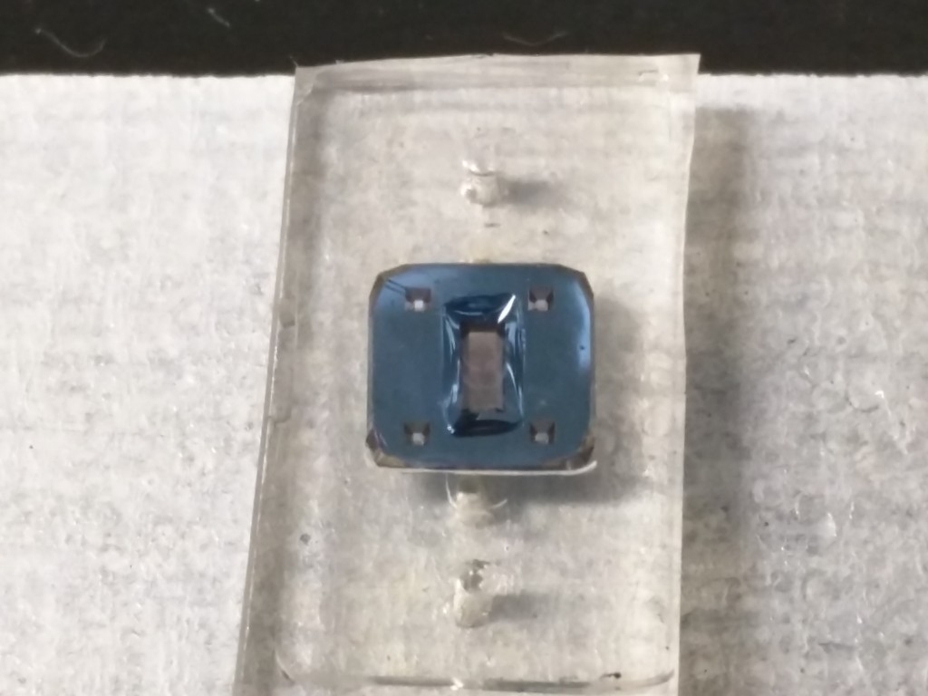
I used a 300 micron gasket as a sealing layer, with a channel cut out in it that exposes only the trench. This also acts as a border and contains the gel inside it. Thus the total thickness of the gel is 300 (silicon substrate) + 300 (gasket) = 600 microns thick. I used this assembled stack to grow endothelial cells on the top and tried to perform neutrophil transmigration through the gel. Similar experiment was performed by Leslie, but using a different gel material, and without endothelial cells. I grew HUVECs on the membrane with geltrex coating above the membrane, and collagen gel below the membrane. After 4 days, I isolated neutrophils and seeded on the endothelial cells. 2 Hours before neutrophil addition, I added fMLP in the basal compartment to a final concentration of 200 nM, so that it can diffuse through the 600 micron gel in the meantime. Both neutrophils and fMLP are in endothelial cell medium, so that ECs remain happy throughout the experiment.
For some reason, I didn’t find the neutrophil transmigration through the gel that impressive. I was going to abandon the experiment, but instead decided to leave it going on inside the incubator. Maybe the fMLP took longer times in collagen gel than in Geltrex gel (as Leslie did). After abut 18 hours, I took the video. Surprisingly endothelial cells were not all dead even in presence of bacterial peptide. Also I was able to record the video involving slicing, showing all the layers of my devices. Here is the first video:
At 10x zoom you can see the endothelial cells and neutrophils (phase bright).
At 20x zoom; you can see only neutrophils; endothelial cells are difficult to image at this magnification.
Summary: I am able to achieve a 3D cell culture environment for cell migration using our membrane in the device configuration. Endothelial cells were not confluent; I need to use flow to align them and grow a tighter monolayer. So neutrophils might as well have went through the open pores rather than going through the endothelial cells. Nonetheless, this capability of adding an external ECM layer brings us one step further in developing a microvessel mimetic in vitro model. More to come soon…