Fluorescent Exosomes Labeled for RNA
This will be a very brief post, but kind of an interesting one. I have been attempting to get my identification of exosomes by immunogold labeling and that hasn’t been going terribly well. I think that it is due to a wealth of complications including a lack of fixed exosomes, salt contamination and other problems. While I fully intend to solve these problems and get that immunogold labeled image, I also wanted to explore alternatives to identifying the exosomes, perhaps one that could be performed without having to dry the membrane. Jim pointed me in the direction of a paper where they used an RNA targeting dye to label exosomes (this paper can be found here) and I followed that direction.
The dye that I used was SYTO RNAselect and it excites at 490 nm, so it is compatible with the FITC or Alexa Fluor cubes. The dye is only supposed to fluoresce when it is bound to RNA, making it a good indicator of being bound inside of exosomes. Therefore, I followed the protocol on the Thermo product page (see the application note here) and labeled some of my previously purified exosomes (the stock that we bought). This led to the following image:
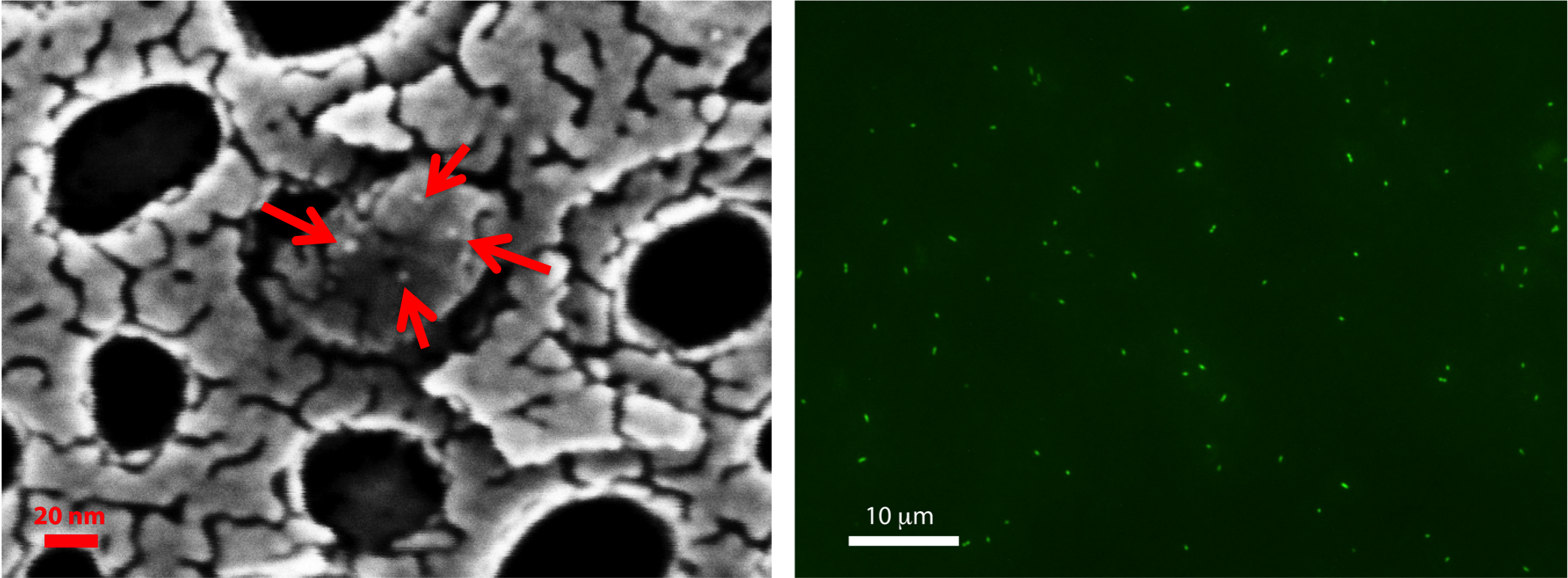
Figure 1: Immunogold (left, anti-CD63) and fluorescently (right, RNAselect) labeled exosomes.
We observed brightly fluorescing dots or clusters of dots that were sub micron, which indicates that these could be exosomes. Their size would appear to be an effect of the fluorescence and I will be running a check with 100 nm beads to confirm that this is how they appear (though Aslan already did experiments with these beads, which can be found in his post here). This is encouraging data and I will next attempt (probably a couple of hours after we go over this) to use detergent to destroy the exosomes and also back wash to try and kick them off.
This is encouraging. I would suggest spiking into plasma a known amount of exosomes labeled with this dye and then running your TF capture protocol. This may show you how well your TF capture protocol does and if plasma somehow complicates the matter. I would also suggest adding RNase to the permeabilized exosomes (e.g. http://www.sigmaaldrich.com/catalog/product/sigma/r4875?lang=en®ion=US).