New Diffusion-Based Separations
Two simple diffusion separations were performed before break using membranes from wafers 205 and 213 (locations unknown as these were salvaged/misplaced chips). I ran the samples I collected on a 10% SDS-PAGE gel today. It seems that the membranes were able to separate the standards at two different cutoffs.
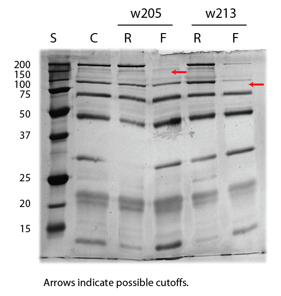
The missing band in the middle of this gel is due to a staining anomaly as the background is also much lighter in that specific area. C = Control, S = Gel Standards, R = Retentate, F = Filtrate. Molecular weights indicated on left side of gel.
Lets get the corresponding pore histograms and see if we can understand the basis for these two separations.
Jess
We discussed that myosin is likely 520k in the natured state and probably 50 nm or larger in one dimension. What about the second protein that is retained by only one of the membranes? What is it, and do we expect that it will be a globular protein at this MW when it is natured?